Prematurity and immature development of the pulmonary system, severe illness, trauma, overdose, aspiration, and other cardiopulmonary conditions call for protection of the airway and establishment of a safe and secure means to ventilate.
By Bill Pruitt, MBA, RRT, CPFT, FAARC
Intubation is needed when respiratory failure has occurred or is anticipated to occur. Prematurity and immature development of the pulmonary system, severe illness, trauma, overdose, aspiration, drowning, and congenital anomalies in the cardiopulmonary system, call for protection of the airway and establishment of a safe and secure means to ventilate. Reintubation may be needed in cases where the endotracheal tube (ETT) has been accidentally removed or dislodged. Intubation in the neonatal and pediatric populations requires special skills and a solid understanding of the anatomy. This article will look at best practices and special considerations for intubation in neonatal and pediatric patients.
Overview of the Anatomy and Physiology
Infants and children undergo changes in the anatomy as they grow, with the differences most pronounced under 2 years of age. Between 2 and 8 years old the airway is in transition, and by age 8 the anatomy is comparable to a small adult.1 Children have several distinctions in the airway anatomy including a larger tongue, a larger and floppy epiglottis, a narrow cricoid ring, and a higher and more anterior glottic opening. At infancy, the glottis is located at C1, moving downward to C3-C5 around age 7, and finally reaching C4-C6.1 The trachea is shorter, more narrow, and more compliant.2 From birth to about 2 years old there is less control of respiration, inefficient use of the muscles of ventilation, different airway and lung mechanics, and a higher basal metabolic rate (increasing the risk of rapid desaturation during intubation). These factors result in higher risk of cardiorespiratory failure, which is even more pronounced during an intubation procedure in neonatal and pediatric patients. The total lung capacity (TLC) and functional residual capacity (FRC) is lower, and the thorax is more compliant while the lungs are less compliant. These issues bring about increased tendency for airtrapping and early closure of the terminal airways.2
Preparing for Intubation
Training for neonatal/pediatric intubation can be done using simulation to allow for a controlled, safe setting, provide time for the team to improve communication, practice using different aids to intubation, and work through different scenarios to prepare for changing conditions or special circumstances. Having an intubation checklist has proven to be very helpful to be sure all the equipment is available and functioning, appropriate medications and respective dosages have been reviewed and prepared, and the intubation team is primed and ready for the procedure. Sample checklists are readily available, and if needed, the team can customize these to fit the particulars of their institution.2-3
Assessment of the patient prior to intubation is needed to evaluate the anatomy and check on the hemodynamic and pulmonary status. Due to the anatomic differences mentioned above, a shoulder roll or folded towel may be useful to position the head and neck so that the vocal cords and glottis can be visualized. Historically, uncuffed ETT have been most commonly used in neonatal patients but use of cuffed tubes are increasing as studies have shown that they may be a better choice.4 Orotracheal intubation is also the most used route but nasotracheal intubation is an available option and may be a consideration as more studies are being published showing favorable outcomes.5
ETT sizes for neonates are often determined by patient weight (2.5 mm tube if <1,000 grams, 3.0 tube if 1,000-2,000 grams, 3.5 mm tube if 2,000-3,000 grams, 3.5 or 4.0 mm tube if >3,000 grams).3 Selecting the right size ETT has been a challenge, with several proposed size prediction models available. An article published in 2022 compared four methods of estimating the correct size tube for pediatric patients and concluded that ultrasonic evaluation of the cricoid cartilage and trachea was the best approach, and using the diameter of the little finger to be the best of the conventional approaches.6
The Intubation Procedure
To intubate, the patient should be properly positioned and preoxygenated. If possible, medications (sedative and paralytic) may be useful to facilitate intubation. Oxygen and bag-mask ventilation should be continued throughout the procedure to avoid desaturation and clear exhaled CO2. Sedation is often accomplished with Fentanyl (4 mcg/kg) followed by saline flush given first, then consider giving Atropine (20 mcg/kg) followed by saline flush to reduce issues with reflex bradycardia and dry secretions. Suxamethonium (2 mg/kg) followed by saline flush is given to paralyze, and the intubation should follow immediately. The procedure should stop if the intubation is not completed within 30 seconds from the time of laryngoscope insertion, if bradycardia <70 beats/minute occurs at any time, or if oxygen saturation drops <70%. Use bag-mask ventilation with oxygen to regain a good baseline before restarting the procedure.3 A bougie catheter may be used to establish the path through the glottis and allow for the ETT to pass over the catheter into the lower airway. Video laryngoscopy may be helpful if the glottis cannot be visualized.6
Correct position in the airway is challenging in neonates and younger pediatric patients due to the short length of the trachea. Prediction models have been used to give guidance for ETT depth but there have been several studies that show problems with the many of the guidelines. Work is continuing to try and improve these models.2,8 Confirmation of ETT placement includes physical examination, auscultation of the chest and epigastrum, capnography, chest radiography, use of light/sound transmitting devices, and ultrasound.2 Movement of the head (by neck flexation or neck extension or turning the head from side to side) moves the ETT up or down in the trachea. This may malposition the ETT so extra care must be taken to insure the tube is at the correct spot and is not malpositioned when procedures or moving the patient (ie transport, holding, repositioning in the bed) occur.2
Securing the Airway
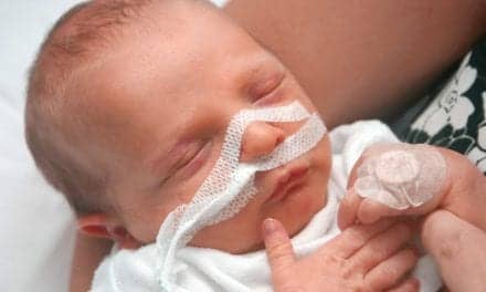
Once an ETT has been inserted and checked for proper position, it must be secured. ETT can be secured by taping or by a tube-holding device. With either approach, extra care must be taken to avoid skin irritation, skin breakdown and pressure sores. If using tape, use of “pre-taping” with a transparent dressing, such as 3M’s Tegaderm, Convatec’s Duoderm OpSite, or 3M’s No Sting barrier film, helps increase tape adhesion and protect the skin. Several tapes are available to use including products such as Beiersdorf Inc’s Leukoplast or Elastoplast, or Kendall Healthcare Products’ WetPruf. It is recommended that benzoin compounds not be used to aid in tape adherence to the skin for securing infant ETTs as damage to the skin may occur when removing the tape.9
Using tape to secure the ETT is difficult in neonates and pediatric patients due to the large amount of oral secretions. Many sites are using ETT holding devices that work effectively in securing the airway. There are a number of devices available on the market, including the AnchorFast ET Tube Holder, Cooper Surgical Neo-fit, Smiths Medical Portex ETTube Holder, Neotech Neo Bar, Ambu ETT Holder, and the Laerdal Thomas Tube Holder.9
COVID-19 Considerations
With the 2020-2021 changes brought on by the COVID-19 pandemic, many procedures had to be evaluated and revised in order to protect the patient, healthcare team, and families. An interim guideline for Basic Life Support (BLS) and Advanced Cardiac Life support (ACLS) for intubation in suspected or confirmed cases of COVID-19 was released in 2022.10 This document emphasizes three main points:
All members of the resuscitation team should wear a respirator (N95), along with other PPE, such as gown, gloves, and eye protection when caring for someone with suspected or confirmed COVID-19 infection. This precaution is empathized for all activities that are consider aerosol-generating procedures (which includes chest compression, defibrillation, bag-mask ventilation, intubation, and positive pressure ventilation).
Resuscitation actions need to be reinforced, as there has been a dramatic decrease in cardiac arrest survival during the COVID-19 pandemic. The cause(s) for this decrease is unclear and complex, but the authors mentioned that there may have been delays in prompt actions due to the added time in donning PPE or in securing the airway. In addition, wearing PPE may have contributed to more rescuer fatigue that brought about a decrease in the quality of CPR being performed.
Take action to have adequate PPE supplies on hand in all clinical settings and stress that all members of the health care team comply with the recommended precautions to avoid possible exposure or spread of the virus. Use of high-efficiency particulate air (HEPA) filters is included in the recommendations to reduce spreading the virus. All ventilation devices (bag-mask, noninvasive and invasive ventilators) need to have HEPA filters in place on the exhaust portion of the device. As an alternative to HEPA filters, a heat and moisture exchanging (HME) filter with >99.99% viral filtration efficiency can be used between the ventilation device and the airway. The HME should remain in place when changing from one device to another (ie switching from bag-mask ventilation to using a noninvasive or invasive ventilation approach).
Video laryngoscopy is recommended to increase the success rate and (possibly) to decrease the time/attempts needed for proper placement of the ETT. Use of a cuffed ETT (rather than uncuffed) is recommended to reduce the potential for aerosolizing respiratory particles. Additionally, the guideline mentions the need to maintain a closed circuit between the ETT and the ventilating devise. With this in mind, endotracheal administration of medications should be avoided due to the need to disconnect from the ventilation source and this may cause unfiltered exhalation produce as aerosol. The guidelines mention that newborn babies are not likely to be a source of COVID-19 transmission even if the mother is confirmed to have the virus, but to keep in mind that the mother will be a potential source of infection (with either confirmed or suspected status).10
Conclusion
Intubation in neonatal and pediatric patients is intense and can be stressful. It requires skill, knowledge, practice, and the right team, right equipment, good communication, and good timing to be successful. These patients cannot be cared for as “little adults” but call for special care due to their size and fragile nature. There is less leeway, less wiggle room for having everything correct; placing the right-sized tube in the right position is more difficult. With the added burden of COVID-19 precautions, this procedure has become more challenging, but a good outcome can still be achieved.
RT
Bill Pruitt, MBA, RRT, CPFT, FAARC, is a writer, lecturer, and consultant. He has over 40 years of experience in respiratory care, and has over 20 years teaching at the University of South Alabama in Cardiorespiratory Care. Now retired from teaching, he continues to provide guest lectures and write. For more info, contact [email protected].
References
- From the Anesthesia Key website: Intubation of the Pediatric Patient | Anesthesia Key (aneskey.com).
- Volsko TA. Kittredge Lecture: Airway Safety in Neonatal and Pediatrics. Respiratory Care. 2022 Jun 1;67(6):756-68.
- Abdelhadi AA, et al. Non-Emergent Endotracheal Intubation of the Newborn: Practical Management. 2022 Mar.
- Chen L, et al. Cuffed versus uncuffed endotracheal tubes in pediatrics: a meta-analysis. Open Medicine. 2018 Jan 1;13(1):366-73.
- Christian CE, et al. Use and outcomes of nasotracheal intubation among patients requiring mechanical ventilation across US PICUs. Ped Crit Care Med. 2020 Mar 11;21(7):620-4.
- Putra SR. Accuracy Comparison between Four Methods of Endotracheal Tube Diameter Estimation for Pediatric Patients: An Observational, Cross-sectional Study.
- Zhou M, et al. Video laryngoscopy improves the success of neonatal tracheal intubation for novices but not for experienced medical staff. Frontiers in Pediatrics. 2020 Aug 6;8:445.
- Volsko TA, et al. Development and internal validation of an equation using anthropometric measures to predict correct endotracheal tube insertion depth. Can J Resp Ther. 2022;58:9.
- Andrews D, et al. Securing paediatric endotracheal tubes: Tape it like you mean it! Australasian Emerg Nurs J. 2007 Mar 1;10(1):30-3.
- Atkins DL, et al. 2022 Interim Guidance to Health Care Providers for Basic and ACLS in Adults, Children, and Neonates With Suspected or Confirmed COVID-19. Circulation: Cardiovascular Quality and Outcomes. 2022 Apr;15(4):e008900.