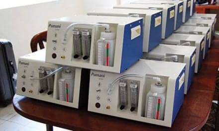
For children with suspected respiratory/metabolic problems, blood gases can provide valuable insights and assist clinicians in developing treatment plans.
By Bill Pruitt, MBA, RRT, CPFT, FAARC
When neonatal and pediatric patients are suspected of having respiratory and/or metabolic problems, blood gases (along with the patient’s history and physical findings) can provide valuable insight into the true condition and guide the treatment choices and plan of care.1-2 Decisions to initiate oxygen therapy, continuous positive airway pressure (CPAP), mechanical ventilation, and use of antibiotics, issues with nutritional support, use of diuretics, etc, may be affected by blood gas results as they are coupled the history and physical exam. This article will look at sampling and analysis of primarily neonatal blood gases and discuss some of the possible conditions associated with abnormal findings in both neonate and pediatric patients.
Samples for Blood Gas Analysis
During and after the delivery of a newborn, blood gas samples can be obtained from the umbilical cord artery or the umbilical vein (by single draw or indwelling line), capillary sampling from heel or finger (or rarely, ear lobe prick), single-draw arterial puncture, or from an indwelling peripheral arterial line in the arm or leg.3 All of these sites are available for use in pediatric patients except the umbilical sites.
Obtaining blood samples from neonates must be done judiciously as multiple samples can reduce their blood volume and bring about hypovolemia.2 Capillary and arterial puncture procedures require that the neonate (or pediatric patient) be carefully immobilized, and that two or more people be present to accomplish this. Crying and struggling before and during this procedure can alter the values (ie, pO2, pCO2) but avoiding these reactions can be a difficult task due to pain and discomfort for the patient.3 Most facilities use a pre-packaged blood gas sample kit that contains all of the components needed to draw the sample. Kits are developed for capillary, arterial puncture, or for drawing a sample from an indwelling catheter. Step-by-step directions for each of these procedures can be found in the WHO guidelines on drawing blood: best practices in phlebotomy (See reference 3).
The single umbilical vein carries oxygenated blood from the mother’s placenta to the fetus while the two umbilical arteries carry blood from the fetus to the placenta that is deoxygenated and full of the carbon dioxide (CO2) eliminated by the fetus through fetal metabolism. Thus, a venous cord blood gas will give insight into the placenta metabolism while the arterial cord blood gas will reflect fetal metabolism at the time of delivery.4 Since veins are more compressible than arteries, when the umbilical cord is compresses venous blood flow is reduced more than arterial blood flow. When this happens, oxygen delivery from the placenta through the cord vein will drop and the fetus responds by extracting more oxygen. This will also cause more CO2 to be eliminated making the arterial cord blood more acidotic. The end result will be an arteriovenous pH difference and samples should be obtained from both the umbilical artery and umbilical vein.4
The 2014 recommendations from the American College of Obstetricians and Gynecologists (ACOG) and the American Academy of Pediatrics call for umbilical cord blood gas analysis to be done in all high-risk deliveries when there is a suspicion of abnormalities in the fetal metabolism (indicated by such issues as abnormal fetal heart rate tracings and low Apgar scores at birth).5 Other indications from the ACOG include cesarean delivery related to fetal compromise, severe growth restriction, maternal thyroid disease, intrapartum fever, or multi-fetal gestation.2 As soon as possible after delivery, a 10 to 20 cm segment of the umbilicus should be clamped and samples should be drawn from the umbilical artery and vein in a pre-heparinized syringe for analysis. pH, pO2 and pCO2 measurements are reliable for up to 60 minutes after birth (with the syringe storage in an ice slurry to reduce ongoing metabolism in the blood) but lactate levels become unreliable around 20 minutes after birth. Therefore it is important to note the source (umbilical artery or vein) and the timing of the sample to inform the interpretation of the results.4
Capillary samples are comparable to arterial blood for pH, pO2, pCO2, and HCO3-. A microsample (100-150 µL) should be taken; often this is done on a prewarmed heel using a prick to allow capillary blood to ooze out and fill a heparinized capillary tube.6 Samples from heel pricks are performed from birth to about 6 months of age, with infant weights from 3 to about 10 kg on the medial or lateral plantar surface. Finger prick samples are performed in patients over 6 months of age, greater than 10 kg, and on the side of the ball of the second or third finger (middle and ring finger).3 The appropriate lancet length should be used to avoid striking a bone or making too deep of a puncture. The first drop of blood should be wiped away as it may be contaminated with tissue fluid or debris from the skin. Squeezing the heel or finger may cause dilution of the specimen with plasma and can increase hemolysis (destruction of the red blood cells), making the results unreliable.3 The heparinized capillary tube usually includes a metal “flea” that moved up and down inside the tube blood sample using a magnetic collar outside the tube to mix the heparin in the blood. Before introducing the sample into the blood gas analyzer, the flea must be removed.6 To avoid air bubbles, the capillary tube should be held horizontal to the sample site.
Single-draw arterial puncture uses a 23-gauge butterfly needle with extension tube connected to a pre-heparinized syringe to obtain the sample. Blood is drawn from the radial artery if possible, but the brachial or femoral arteries may also be accessed. Issues with the alternate sites include the fact that they are harder to locate than the radial artery (being less superficial), have less collateral circulation, and are close to structures that could be damaged by faulty technique.3
Drawing arterial blood gas (ABG) from an indwelling arterial catheter avoids causing discomfort to the neonate or pediatric patient and also avoids the potential alteration of results that can be brought on by crying/struggling. Due to issues with possible thrombosis and infection, arterial catheters are used in only in critically ill patients. Arterial catheter samples may have issues with hemodilution due to the use of heparinized saline solution used to perfuse the site. If the catheter is not adequately cleared of the saline solution, the sample results will have lower pCO2, and HCO3– values due to the hemodilution.1
Table 1a. Normal Neonatal ABG Values6
| pH | 7.35 to 7.45 |
| paCO2 | 35 to 45 mm Hg |
| paO2 | 50 to 70 mm Hg |
| HCO3– | 20 to 24 mEq/L |
| BE | +/- 5 |
Table 1b. ABG Values Based on Neonatal Age6
| Pre-birth (scalp) | 5 min after birth | 1-7 days after birth | |
| pH | >7.20 | 7.20 to 7.34 | 7.35 to 7.45 |
| pCO2 | <50 mmHg | 35 to 45 mmHg | 35 to 45 mmHg |
| pO2 | 25 to 40 mmHg | 49 to 73 mmHg | 70 to 75mmHg |
| Sat% | >50 | >80 | >90 |
| HCO3– | >15 mEq/L | 16 to 19 mEq/L | 20 to 24 mEq/L |
Analysis of Blood Gas Samples
Blood gas samples can be analyzed at bedside or other non-centralized location using small, handheld point-of-care (POC) or portable analyzers on a cart, or on larger non-portable analyzers (usually located in a centralized lab). There are many blood gas analyzers on the market for both POC/portable and centralized locations. Care in obtaining the sample is required to avoid issues with air bubbles or delays in running the sample as these will alter the results. Depending on the particulars of the machine, blood gas analyzers can provide a variety of results including: pH, pCO2, pO2, Na+, K+, Ca++, Cl–, Glucose, Lactose, Hct, tHb, OvHb, COHb, MetHb, HHb, tBili, sO2, BE(B), BE(ecf), tHb(c), Ca++ (7.4), Anion gap (AG), P/F ratio, pAO2, CaO2, CvO2, p50, O2cap, sO2(c), O2ct, HCO3 – std, TCO2, HCO3– (c), A-aDO2, paO2/pAO2, RI, CcO2, a-vDO2, Qsp/Qt (est), Qsp/Qt, Hct(c), and more.
Interpretation of Blood Gases
For pediatric patients and babies that are approaching 1 month of age, interpretation of arterial blood gases (ABGs) is fairly straightforward and follows the usual pathways for understanding the results. The normal ranges for the major components are: pH 7.35-7.45, PaO2 80-100 mmHg, PaCO2 35-45 mmHg, HCO3– 22-26 mEq/L.2,7 The four steps to interpretation in this setting are:
Step 1. Does the pH reflect an acidosis or alkalosis?
Is the pH above 7.40 (moving to an alkalosis) or less than 7.40 (moving to an acidosis). If pH is still within the normal range but either the PaCO2 and/or the HCO2– will be out of their respective normal ranges and the ABG could be either uncompensated or partially compensated (see Step 3).
Step 2: For either an acidosis or alkalosis, what is the cause?
- For a respiratory alkalosis the PaCO2 will be low (<35 mmHg) with a high pH.
- For a respiratory acidosis the PaCO2 will be high (>45 mmHg) with a low pH.
- For a metabolic alkalosis the HCO3– will be high (>26 mEq/L) with a high pH.
- For a metabolic acidosis the HCO3– will be low (<22 mEq/L) with a low pH.
- For a combined alkalosis (respiratory and metabolic) the PaCO2 will be low and the HCO3– will be high (both out of their respective normal ranges) with a high pH
- For a combined acidosis (respiratory and metabolic) the PaCO2 will be high and the HCO3– will be low (both out of their respective normal ranges) with a low pH.
Step 3: Is there compensation occurring to correct the problem?
Compensation occurs as the body tries to bring the pH back toward or into the normal range. If the respiratory system is the cause, the metabolic system compensates by changing the HCO3– in the opposite direction (ie, a respiratory acidosis will bring about a rising HCO3-). This is a slow process, occurring over a number of days. When full compensation is achieved, the pH will be in the normal range and both the PaCO2 and the HCO3– will outside their normal ranges but in opposite conditions (one being alkalotic while the other is acidotic). If the problem is a combined condition, there is no compensation occurring as both the respiratory and metabolic systems are contributing to the issue. In partial compensation,
the PaCO2 and the HCO3– will outside their normal ranges and in opposite conditions, but the pH will still be outside the normal range. For uncompensated situations, the pH and the system causing the problem will be out of their normal ranges and the other system (metabolic or respiratory) will be within its normal range.
Step 4: Is there a problem with oxygenation?
If the PaO2 is below the normal range, hypoxemia is occurring. If the PaO2 is above the normal range, hyperoxia is occurring. Either of these situations can cause problems and the abnormality needs to be corrected (see reference 8 for more details on interpretation).8
For neonates, blood gases will be rapidly changing as the newborn converts from fetal circulation to autonomous circulation. At the same time gas exchange begins as the newborn starts breathing and the lungs take over what had been the task of the mother’s placenta. According to a 2002 article by Fouse: “The placenta acts both as “lungs” and “kidneys” for the fetus by supplying oxygen and removing carbon dioxide and metabolites.2 It provides efficient gas exchange as well as allows nutritional substances such as vitamins, glucose, free fatty acids, and electrolytes to pass between the two circulations without allowing the two circulations to mix.”9 At birth, PaO2 and pH rapidly climb from low to normal while PaCO2 rapidly drops from high to normal (most of this occurs within 30 minutes after delivery if all systems are functioning correctly).1
Mean arterial umbilical cord blood gases in neonates (with uncomplicated delivery) will have a pH around 7.24 to 7.27 and the venous cord blood gas will have a pH around 7.32 to 7.34. Cord blood gases from the umbilical artery will have a mean PaO2 of 31.5 mmHg while a cord blood gas from the umbilical vein will have a mean PaO2 of 43.5 mmHg (reflecting the mother’s respiration). Contractions during labor cause metabolic stress and a lower cord pH, while a neonate who is delivered by caesarian section (not subjected to labor) will have a higher cord pH.4 See Table 3 for common causes of acid-base abnormalities in neonates.10
Capillary blood gases provide values for pH and PCO2 that are very close to what would be seen with an arterial blood gas but are not very reliable in cases of hypotension, poor perfusion, or cold peripheries. Capillary blood gases are also not reliable for assessing oxygenation status or for assessing hypoxemia.11-12 Assessment of oxygenation relies on an arterial sample.
Table 3. Common Causes of Acid-Base Abnormalities in Neonates
| Respiratory acidosis | • Upper airway obstruction, choanal atresia, Pierre-Robin • Lung abnormalities: respiratory distress syndrome, transient tachypnea of newborn, pneumonia, meconium aspiration, pulmonary hypoplasia, congenital malformations • Central hypoventilation syndrome: encephalopathy, hemorrhage • Neuromuscular insufficiency: congenital myopathies, congenital neuropathies • Iatrogenic: inadequate ventilator settings |
| Metabolic acidosis | With increased anion gap • Lactic acidosis: sepsis, hypoperfusion, shock, necrotizing enterocolitis • Inorganic acidosis: inborn errors of metabolism, renal failure With normal anion gap • Increased gastrointestinal H2CO3 loss (ileostomy, diarrhea) • Renal tubular acidosis |
| Respiratory alkalosis | • Iatrogenic: excessive ventilation • Compensatory hyperventilation: hypoxic-ischemic encephalopathy |
| Metabolic alkalosis | • Gastric losses: vomiting, non-replacement of excessive nasogastric aspirates • Thiazide and loop diuretics • Hypokalemia |
Conclusion
Blood gases in the neonatal and pediatric populations are valuable in assessing acid-base status and ventilation, and depending on the source of the blood, oxygenation. Neonatal blood gases must be carefully interpreted and consider the time of the sample as conditions are rapidly changing from delivery to early life independent from the mother. If a prick or a stick is required, the patient’s reaction to pain (struggling, crying) can cause changes from the baseline in the results, so these factors need to be considered in interpreting the blood gases.
Point-of-care testing can bring faster results compared to tests run in a centralized location, but the cost of these analyzers and the necessary supply costs may be a factor in using this approach and this may impact the widespread use. Respiratory therapists are essential to obtaining and running the samples, and also integral to the actions taken in response to abnormal blood gases—so a sound and thorough understanding of neonatal/pediatric blood gases is paramount in those who work in the neonatal and pediatric areas.
RT
Bill Pruitt, MBA, RRT, CPFT, FAARC, is a writer, lecturer, and consultant. He has over 40 years of experience in respiratory care, and has over 20 years teaching at the University of South Alabama in Cardiorespiratory Care. Now retired from teaching, he continues to provide guest lectures and write. For more info, contact [email protected].
References
- Brouillette RT, Waxman DH. Evaluation of the newborn’s blood gas status. Clinical Chemistry. 1997 Jan 1;43(1):215-21.
- Mulligan M. From the AcuteCareTesting .org website. “Blood gas interpretation in the neonate – what do you need to know now?”: https://acutecaretesting.org/en/articles/blood-gas-interpretation-in-the-neonate. Accessed Jan. 2023.
- Dhingra N, Diepart M, Dziekan G, Khamassi S, Otaiza F, Wilburn S. WHO guidelines on drawing blood: best practices in phlebotomy. From Geneva: World Health Organization; 2010. 2, Best practices in phlebotomy. Available from: https://www.ncbi.nlm.nih.gov/books/NBK138665/
- Saneh H, Mendwz M, Srinivasan V. “Cord Blood Gas” from the StatPearls website: https://www.ncbi.nlm.nih.gov/books/NBK545290/. Accessed January 2023.
- Executive summary: Neonatal encephalopathy and neurologic outcome, second edition. Report of the American College of Obstetricians and Gynecologists’ Task Force on Neonatal Encephalopathy. Obstet Gynecol. 2014 Apr;123(4):896-901.
- Deorari A. From the All India Institute Of Medical Sciences (AIIMS) https://newbornwhocc.org/pdf/Blood-Gas-Book-workbook-2008.pdf. Accessed Jan. 2023.
- From the University of Iowa “Pediatric Reference Ranges” website: https://www.healthcare.uiowa.edu/path_handbook/Appendix/Heme/PEDIATRIC_NORMALS.html. Accessed Jan. 2023
- Pruitt B. “A Discussion of Arterial Blood Gas Analysis and Interpretation” from RT for Decision-makers website 2022: https://respiratory-therapy.com/products-treatment/diagnostics-testing/diagnostics/discussion-arterial-blood-gas-analysis-interpretation/. Accessed Jan. 2023.
- Fouse B. From AcuteCareTesting.org website. “Reference range evaluation for cord blood gas parameters” https://acutecaretesting.org/en/articles/reference-range-evaluation-for-cord-blood-gas-parameters.
- Tan S, Campbell M. Acid–base physiology and blood gas interpretation in the neonate. Paediatrics and Child Health. 2008 Apr 1;18(4):172-7.
- Goel N, Calvert J. Understanding blood gases/acid–base balance. Paediatrics and Child Health. 2012 Apr 1;22(4):142-8.
- Martin R, Deakins K. “Respiratory support, oxygen delivery, and oxygen monitoring in the newborn” from the UpToDate website: https://www.uptodate.com/contents/search. Accessed January 2023.


