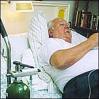
From its developmental stages to its future of signal extraction, pulse oximetry provides an increasingly accurate, noninvasive method of collecting ABG analysis data.
Often referred to as the fifth vital sign, pulse oximetry has gained rapid and widespread acceptance among clinicians from all disciplines. Once solely the domain of clinical pulmonary function laboratories, pulse oximetry and its predecessor ear oximetry have evolved into a ubiquitous technology.
Pulse oximetry’s presence in operating suites, recovery rooms, intensive care units, delivery rooms, nurseries, emergency departments, emergency vehicles, general care areas, home care, practice offices, and many other areas cannot be denied. No one would argue that patient care has not been improved through the use of pulse oximetry. It is easy to use, affordable, accepted by insurance companies for reimbursement, and noninvasive. What more could one ask of a technology?
Gone are the days when arterial blood gas (ABG) specimens were drawn for every change made to ventilator parameters or for every change in the patient’s fraction of inspired oxygen. This is not to say that ABG analysis is obsolete, by any means, but it is invasive and not a continuous real-time measurement. For years, the search for accurate, inexpensive, and noninvasive methods for collecting the data ABG analyses offer continued. Pulse oximetry is one technology that has allowed great strides in noninvasive monitoring, but there is still much work to do.
Obtaining accurate pulse-oximetry readings in motion and low perfusion states, the development of a central sensor probe, miniaturization, and wireless technology are the future focus points for the ongoing development of pulse oximetry (as well as of noninvasive carbon-dioxide monitoring and pH monitoring). The less invasive clinical practices are, the more patients will benefit.
Pulse Oximetry’s Development
Pulse oximetry is not new. Based on the Lambert-Beer relationship between light transmission and optical density (known as Beer’s law), the developmental phase of pulse oximetry can be traced back to the late 1800s. The development of the spectrometer by Bunsen and Kirchhoff in 1860 and the demonstration of the oxygen-transport function of hemoglobin by Stokes and Hoppe-Seyler shortly thereafter paved the way for the development of pulse oximetry.
By 1932, scientists such as Nicolai (who measured in vivo oxygen consumption) and Kramer (who measured saturation by the transmission of light through unopened arteries) moved closer to pulse oximetry. In 1935, Matthes developed the first two-wavelength ear oxygen-saturation probe. The device worked, but it was slow, required frequent calibration, needed bulky equipment, and showed poor differentiation between arterial and venous blood. The original device used a light source balanced with red and green filters. Later, the accuracy of the device was improved by switching to red and infrared filters. Infrared filtration is the basis of today’s pulse oximetry.1
In 1942, Millikan used a heated ear probe to determine why fighter pilots blacked out under high gravitational forces. Millikan is credited with coining the term oximeter. The device featured an ear clip connected to onboard recorders in the aircraft. Later, in 1949, Wood redesigned the device to include a pressure capsule; this squeezed blood out of the ear to obtain an absolute zero to improve the accuracy of the device. As the pressure was released and blood was allowed to reperfuse the sample site, the difference between the zero point and the peak was measured, giving the actual saturation. The device, however, suffered from the use of unstable photocells and light sources, and it was never used clinically.2
In 1964, Shaw assembled the first absolute-reading ear oximeter by using a spectrum of eight wavelengths of light. The device worked and was marketed, but its use was limited to pulmonary function laboratories due to its cost and size. In 1972, Aoyagi developed conventional pulse oximetry using the ratio of red to infrared wavelengths of light through the pulsating components of the perfusion cycle (assumed to be only arterial blood) within the measurement area. In this way, he was able to calculate oxygen saturation without calibration.
In 1981, this technology was the subject of commercial development and distribution. Smaller probes were developed that used light-emitting diodes. Although the new probes and monitors were accurate in steady-state situations, motion artifact, low perfusion, electrocautery interference, and ambient light limited the effectiveness of conventional pulse oximetry.3
Present Technology
Current clinical practice remains at this level, but a new technology for signal extraction has emerged; it uses proprietary algorithms (coupled with adaptive filter technology) to separate the arterial signal from nonarterial noise (venous blood movement). The result is a pulse-oximetry technology that has been scientifically and clinically proven to be accurate during patient motion and low perfusion in adults and neonates, and able to deliver superior performance in states of unfavorable ambient light, and electrical noise.4,5 Currently, this technology is being licensed to several pulse-oximeter manufacturers.
Future Prospects
Much work that focuses on obtaining accurate pulse-oximetry readings in low perfusion states and during movement is now in progress, as are efforts to miniaturize pulse oximeters and adapt them for wireless data transmission. To that end, one of the strongest and most innovative technologies supporting pulse-oximetry accuracy is signal extraction.
The major difference between conventional pulse-oximetry technology and signal extraction is the concept of physical separation of arterial signals and venous signals. In conventional pulse oximetry, the measured values are assumed to be arterial.
In conventional pulse oximetry, red and infrared wavelengths of light are used to measure arterial blood’s oxygen saturation by analyzing the resulting red-infrared ratio. This ratio is, in turn, calibrated to produce a reading of the percentage of oxygen saturation. In order to maintain accuracy, however, a steady-state environment and strong signal distinction are required. Movement affects the reading obtained; in addition, anything that would affect signal strength (such as fluctuating perfusion or poor emitter-to-detector alignment) can also cause erroneous or unstable results.
These artifacts, taken together, constitute the noise component of the signal-to-noise ratio. Signal extraction begins with the knowledge that, during patient motion, the low-pressure venous system is more susceptible to perfusion changes (due to compression and deformity of the vascular bed) than the arterial system. Hence, the venous system is a significant source of noise in the measured signal.
Through the use of an adaptive noise canceller, this venous noise can be filtered out, leaving, in effect, a stronger arterial signal. This allows accurate calculation of optical density ratios. The result is a more accurate and stable oxygen saturation value.
Conclusion
Pulse oximetry is truly vital to medical care, but this prominence carries a responsibility. Too often, pulse oximetry readings are misinterpreted or are erroneous for various reasons (see sidebar). The tendency to accept the percentage at face value is high. Many expensive probes continue to be discarded prematurely as defective. Direct patient interventions can also occur based on erroneous results; worse, patients can be inappropriately monitored, giving caregivers a false sense of security. Pulse oximetry has come a long way since its inception in the early 1800s and with future technology, pulse oximeters will continue to improve patient care more effectively and efficiently.
Gary T. Rushworth, RRT, is a critical care respiratory therapist at Greater Baltimore Medical Center, Baltimore.
References
1. Severinghaus JW, Astrup PB. History of blood gas analysis. VI. Oximetry. J Clin Monit. 1986;2:270-288.
2. Severinhaus JW, Astrup PB, Murray JF. Blood gas analysis in critical care medicine. Am J Respir Crit Care Med. 1998;4:S122.
3. Hough SW. Pulse oximetry. 1997. Available at http://www.mghdacc.com/BaefFolder/PulseRaw.htm.
4. Barker SJ, Novak S, Morgan S. The effects of motion on the performance of pulse oximeters in volunteers. Anesthesiology. 1997;86:101-108.
5. Barker SJ, Novak S, Morgan S. The performance of three pulse oximeters during low perfusion in volunteers. Anesthesiology. 1997;87:A409.


