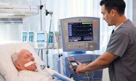
Bronchoscopy plays a role in both diagnosis and treatment of lung disease and recent advances in the procedure have greatly enhanced the usefulness of the fiberoptic bronchoscope (FOB).
By Bill Pruitt, MBA, RRT, CPFT, AE-C, FAARC
Diagnosis of lung disease often requires a combination of tests or examinations to reach a firm conclusion while ruling out other possible issues. Therapies to treat lung diseases (or relieve symptoms) may call for combinations of approaches (ie, physiotherapy, pharmacologics, and surgical or bronchoscopic procedures). Bronchoscopy plays a role in both diagnosis and treatment of lung disease and recent advances in the procedure have greatly enhanced the usefulness of the FOB. This article will discuss the use of bronchoscopy, both as a diagnostic tool and an airway clearance therapy for patients with acute or chronic lung conditions such as bronchiectasis, COPD, interstitial lung diseases, cancer, airway obstruction, and infections.
Bronchoscopy: An Overview
The first bronchoscopic procedure was done in 1897 to remove a foreign body using a rigid bronchoscope by a Gustav Killian—a German physician referred to as the “father of bronchoscopy.”1 The idea for a flexible fiberoptic bronchoscope (FOB) was developed in 1962 by Shigeto Ikeda, a Japanese thoracic surgeon. The FOB became available in 1966, followed closely by the first video bronchoscope in 1967.1 The rigid scope is still used today to perform procedures in the large airways (retrieve foreign bodies, relieve massive airway bleeding, obtain biopsy specimens, dilate airway strictures, insert stents, perform laser therapy, etc).2 The smaller, flexible FOB allowed access to distal airways and advances in bronchoscopy procedures have brought many changes, from obtaining specimens through transbronchial needle aspiration biopsy (TBNA) in the early years to placing airway stents to using
endobronchial ultrasound, electromagnetic navigation tools, robotic bronchoscopy, and more.3 The significant increase in procedures, techniques, navigational tools, etc, has enhanced the term “interventional bronchoscopy” used to describe these special approaches in pulmonary medicine.
Therapeutic Bronchoscopy
Therapeutic bronchoscopy involves using the bronchoscope to treat lung disease. These procedures are used to address issues in asthma, emphysema, COPD, bronchiectasis, lung cancer, removal of foreign bodies, hemoptysis and/or copious secretions, and more. Rigid bronchoscopes are utilized when a larger working channel is needed, as in clearing massive hemoptysis, relief of central airway obstruction due to cancer or other tissues impinging the airway, removal of clots or mucus plugs, or large foreign bodies. Rigid bronchoscopes are utilized in the operating room with the patient under general anesthesia and are more often used in treatment/therapy versus diagnosis.4
Asthma
Bronchial thermoplasty in asthma uses a FOB to deliver thermal energy to the airway walls, which results in a reduction of smooth muscle and reduces the impact of bronchospasm in hyperactive airways. Research outcomes of bronchial thermoplasty include reduction in exacerbations and emergency department visits, improved peak expiratory flow, improvement in scores on asthma control questionnaires, more symptom-free days, reduced medications (both rescue medications and corticosteroids).5 The 2020 Global Initiative for Asthma guidelines recommends bronchial thermoplasty as a potential treatment option for patients at Step 5 with uncontrolled asthma despite optimized treatment regimes.6
Emphysema
In patients with severe emphysema (flattened diaphragm, greatly increased FRC and RV with airtrapping and hyperinflation), the FOB is being used to reduce hyperinflation by three different techniques including endobronchial valves, coils, and thermal vapor ablation. Endobronchial valves (EBVs) are one-way valves that are positioned by bronchoscopic insertion into the segmental bronchi of a hyperinflated lobe. The valves allow for expiratory flow only so that eventually the targeted lobe fully deflates. This relieves the stress on other normal lung tissue by reducing hyperinflation and decreases the FRC and RV.5 Coils are also placed in the airway and designed to gather and compress damaged lung tissue, reduce airway collapse and air trapping, increase lung recoil to enhance exhalation, and redirect air to healthier portions of the lung. The procedure is done via FOB and about 10 coils of different sizes are placed in the targeted area.5 Outcomes observed included improvement in the St George’s Respiratory Questionnaire (SGRQ), and in FEV1, RV and the 6-minute walk test (6MWT).5 Research using randomized clinical trials regarding coils also noted that pneumonia and pneumothorax were the two main serious adverse events, and that “risks of pneumonia and pneumothorax require prespecified therapeutic management plans.”5
The thermal vapor ablation procedure (not approved in the US) uses a FOB to introduce heated water vapor into the upper lobes targeting the areas of blebs and bullae where significant alveolar damage (with airtrapping and increased deadspace) has occurred. The heated vapor causes an inflammatory response leading to scarring of the parenchyma resulting in lung volume reduction (reduced FRC and RV).5 Research outcomes include increased FEV1 and SGRQ. The 2020 Global Initiative for Chronic Obstructive Lung Disease (GOLD) report recommends consideration of endobronchial valves, coils, or thermal ablation in emphysema to reduce significant refractory hyperinflation in patients with severe emphysema.7
COPD
COPD may also be addressed in a bronchoscopic procedure using targeted radiofrequency energy that disrupts the vagal impulses and decreases the parasympathetic overactivity associated with COPD—particularly in the chronic bronchitis phenotype. Described as targeted lung denervation (TLD), the end result is a reduction in the release of acetylcholine with subsequent reduction in smooth muscle constriction.5,8 (This approach has a similar effect as various parasympatholytic or anticholinergic medications such as atropine, ipratropium bromide, tiotropium bromide, and glycopyrrolate but TLD is a physical change with much longer lasting effects). Post hoc analysis of research in TLD has shown that the combination of TLD with long-acting parasymptholytic medications has a synergistic effect compared to TLD alone—this needs further investigation.8
Procedures to destroy hyperplastic goblet cells and reduce the excess mucus production in chronic bronchitis are also being researched. Use of liquid nitrogen, bronchial thermopasty, and balloon disruption/destruction of goblet cells are being investigated. If data supports usage these may be added to the bronchoscopy approach to this chronic disease. Excessive mucus production is mostly found in the goblet cells found in the airways down to the fifth generation and this area of the lower airway is well within the reach of the FOB for possible therapeutic approaches.8
Non-cystic Fibrosis Bronchiectasis
In patients with non-cystic fibrosis bronchiectasis, persistent cough and large amounts of purulent sputum is frequently a major issue. Bronchoscopy has been shown to be effective in airway clearance through bronchoalveolar lavage (BAL) and suctioning to remove retained secretions. A recently published randomized control trial looked at using BAL to manage hospitalized patients with acute exacerbation of bronchiectasis. In this novel study, researchers used normal saline (total amounts ranging from 120mL to 200mL) to lavage and suction these patients and compared their course to a control group of similar patients. BAL therapy significantly increased the time to first acute exacerbation post-discharge and improved COPD Assessment Test (CAT) and Leicester Cough Questionnaire (LCQ) scores.9 The subgroup of patients who had the most benefit from BAL were those with severe bronchiectasis with Bronchiectasis Severity Index (BSI) scores >8, two or more acute exacerbations in the last year, SGRQ >15, CAT >11, or LCQ <16.9
Lung Cancer
The role of bronchoscopy in patients with lung cancer has many facets including diagnosis and treatment. These start with improving the specific diagnosis by obtaining tissue samples with enhanced capability to reach more remote areas through use of endobronchial ultrasound and electromagnetic navigation tools. Therapeutic procedures include various ablation techniques to kill cancer cells (laser, radiofrequency, thermal ablation, electrocoagulation, argon plasma coagulation, cryoablation to freeze cells).10-11 Other bronchoscopic approaches use radioactive pellets (endobronchial brachytherapy) to destroy cancerous cells, or the procedure may attack cancer cells by local injection of chemotherapeutic agents or therapeutic genes. Finally, balloon dilation and stenting has also been used in treatment to improve severe stenosis of the trachea and the main bronchi.10
Diagnostic Bronchoscopy
Diagnosis of infectious lung disease (including bacterial, viral and fungal) and cancer by bronchoscopy has been well established and recent additions to the bronchoscopic procedure have greatly enhanced the diagnostic capabilities. Combining high-resolution computerized tomography (HRCT) with navigational technology (electromagnetic navigational bronchoscopy, ENB) has allowed for collection of more precise and more distal cellular samples and increased the areas that can be reached and treated by bronchoscopy.12 At the same time as these technological advances were coming on board, endobronchial ultrasound (EBUS) also allowed for improved guidance to areas deep in the lung and improved obtaining cell sample. Virtual bronchoscopic navigation (VBN) uses software to combine HRCT data, FOB location in the airways, and the FOB images to create a real-time virtual image to allow for targeting specific areas of concern, improved localization of lesions, and increase collection of needed tissue/cell samples for diagnosis. Development of ultrathin FOBs with an outer diameter of around 2.8 to 3.5mm have contributed to the greater “reach” in obtaining tissue or cell samples in small airways. Lastly, robotic-assisted bronchoscopic systems have helped navigate to small, peripheral airways and allow for precise targeting for obtaining diagnostic samples. (ENB, EBUS, VBN, ultrathin scopes and use of robotic bronchoscopy have also aided in advancing therapeutic procedures such as cryotherapy, ablation, laser vaporization, stent placement, etc, mentioned earlier).12
COVID-19 and Bronchoscopy
Patients with COVID-19 and receiving mechanical ventilation are at risk of having additional infectious agents according to recent research in this area. Researchers looked at 179 patients and used 389 BAL samples obtained by FOB to investigate this and found that 21% of patients in samples obtained early in the course had bacterial superinfections, which were mainly caused by antibiotic-sensitive strains of Streptococcus species and methicillin-sensitive S. aureus. Late BAL samples found ventilator-associated pneumonia in 44% of patients. This resulted in adjustments in the antibiotic regime being used for these patients.13 In this paper from 2021, “The authors detected the pathogens because they looked hard to find them.” In the limitations, the authors noted, “The current analysis did not provide data on fungal superinfections, which is an area of major diagnostic uncertainty and active investigation.”13 Bronchoscopy has been cited as a risky procedure due to the generation of aerosols and increased exposure to the bronchoscopy team, but in this research, the authors found that the procedure could be performed safely if done with all precautions in place and that hospitals “should not be dissuaded from performing BAL when clinically indicated.”13 A recent publication on bronchoscopy in COVID-19 patients in the ICU states, “In general, bronchoscopy has not shown any definitive increase in transmission when proper precautions have been observed”14 and “The risks of periprocedural complications and SARS-CoV-2 transmission among HCWs during bronchoscopy appear to be low. An outbreak is unlikely if appropriate safety measures are followed.”15
Conclusion
Interventional bronchoscopy has significantly changed and enhanced the role of the bronchoscopist and the team that works alongside. Many of these procedures are much longer and require hours of work by the physician, respiratory therapist, nurses, radiology team members, and others to accomplish the job. In COVID-19 patients, FOB is being utilized more often as the early warnings about possible risk of transmission is fading. According to a 2020 AJRCCM article, “In the near future, new approaches for many different lung diseases should become available: biodegradable stents, second- and third-generation EBVs, better nonpharmacological treatments for chronic bronchitis and airflow obstruction, and new treatments in patients with emphysema who exhibit [collateral ventilation].”12 The article continued: “With new options, new uses for interventional bronchoscopy are emerging, and it is plausible that interventional pulmonology has enormous potential to provide safe and effective diagnostic and therapeutic procedures at reduced costs for many patients with a variety of lung disorders.”12
RT
Bill Pruitt, MBA, RRT, CPFT, AE-C, FAARC, is a writer, lecturer, and consultant. He has over 40 years of experience in respiratory care, including more than 20 years teaching at the Univ of South Alabama. Now retired from teaching, he continues to provide guest lectures and write. For more information, contact [email protected]. Full references at www.respiratory-therapy.com.
References
- From Thoracic Key website: https://thoracickey.com/history-of-bronchoscopy-the-evolution-of-interventional-pulmonology/. Accessed 1/3/22.
- From Operative Techniques website: https://www.optechtcs.com/article/S1522-2942(12)00052-9/fulltext . Accessed 1/3/22.
- Panchabhai TS, Mehta AC. Historical perspectives of bronchoscopy. Connecting the dots. Annals of the American Thoracic Society. 2015 May;12(5):631-41.
- From the Mulitmedia Manual of Cardiothoracic Surgery – Operative rigid bronchoscopy: indications, basic techniques and results. https://mmcts.org/tutorial/47. Accessed 1/5/22.
- Perotin JM, Dewolf M, Launois C, Dormoy V, Deslee G. Bronchoscopic management of asthma, COPD and emphysema. European Respiratory Review. 2021 Mar 31;30(159).
- From the 2020 Global Initiative for Asthma guidelines website: https://ginasthma.org/wp-content/uploads/2020/04/GINA-2020-full-report_-final-_wms.pdf. Accessed 1/4/22.
- From the 2020 Global Initiative for Chronic Obstructive Lung Disease (GOLD) website: https://goldcopd.org/gold-reports/. Accessed 1/4/22.
- Hartman JE, Garner JL, Shah PL, Slebos DJ. New bronchoscopic treatment modalities for patients with chronic bronchitis. European Respiratory Review. 2021 Mar 31;30(159).
- Liu Y, Lu HW, Gu SY, Wang WW, Ge J, et al. Bronchoscopic airway clearance therapy for acute exacerbations of bronchiectasis. EBioMedicine. 2021 Oct 1;72:103587.
- Wang Y, Chen E. Interventional bronchoscopic treatment of lung cancer. Laparoscopic, Endoscopic and Robotic Surgery. 2021 Oct 7.
- Mondoni M, Rinaldo RF, Carlucci P, Terraneo S, Saderi L, et al. Bronchoscopic sampling techniques in the era of technological bronchoscopy. Pulmonology. 2020 Jul 2.
- Criner GJ, Eberhardt R, Fernandez-Bussy S, Gompelmann D, Maldonado F, et al. Interventional Bronchoscopy. American journal of respiratory and critical care medicine. 2020 Jul 1;202(1):29-50.
- Kitsios GD, Morris A. Seek and Ye Shall Find: COVID-19 and Bacterial Superinfection. American Journal of Respiratory and Critical Care Medicine. 2021 Oct 15;204(8):875-7.
- Nitesh J, Kashyap R, Surani SR. What we learned in the past year in managing our COVID-19 patients in intensive care units?. World Journal of Critical Care Medicine. 2021 Jul 9;10(4):81.
- Saha BK, Chaudhary R, Saha S, Bonnier A, Chong WH, Chenna P. Bronchoscopy During Coronavirus Disease 2019 Pandemic: A Bronchoscopist’s Perspective. Critical Care Explorations. 2021 Sep;3(9).