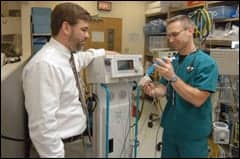
A 57-year-old female patient with severe bullous emphysema benefited from a pulmonary rehabilitation program before and after lung transplantation.
 There was a time when pulmonary rehabilitation was considered only when all else failed in the treatment of chronic lung disease. It was typical for physicians to tell their patients that nothing more could be done for them, so they should try pulmonary rehabilitation. With their medication options exhausted, these patients reported for rehabilitation sessions that they saw as only one step short of going home to die.
A dramatic shift in that attitude has been seen in the 1990s. Pulmonary rehabilitation has enjoyed a transformation from last resort to standard of care. With advances in research, improvements in therapy, and changes in attitude, pulmonary rehabilitation is now accepted as a part of initial treatment.1 It is the constant in the lung-volume–reduction trial (National Emphysema Treatment Trial [NETT]), with all candidates receiving rehabilitation and the two randomization factors being the use of medication alone or of surgical intervention.2
Those conducting outpatient pulmonary rehabilitation programs see an entire spectrum of breathing problems, from chronic obstructive lung disease to fibrosis to pneumoconiosis to sarcoidosis to instances of diaphragmatic paralysis and postpolio syndrome. The programs review the proper use of medications and oxygen and teach energy conservation and effective breathing techniques. They provide the exercise, education, and socialization intended to improve not only the participants’ physical health, but also their quality of life.
Different programs approach that challenge in different ways. Nationwide and worldwide, one can find RCPs, exercise physiologists, registered nurses, and physical therapists heading pulmonary rehabilitation programs. A 1994 definition of pulmonary rehabilitation from the National Institutes of Health cites the “multidimensional continuum of services directed by an interdisciplinary team of specialists.”3 That interdisciplinary approach includes RCPs, physical and occupational therapists, dietitians, social workers, and psychologists, all working under the medical direction of pulmonologists and other medical specialists.
The effectiveness of each of the various components of pulmonary rehabilitation, including upper-extremity and lower-extremity training, ventilatory muscle training, and the psychosocial, behavioral, and educational components and outcomes of programs, was examined in a special report issued in 1997.4
Exercise prescription policies may vary. Some programs set target heart rates using exercise-testing results from the admission evaluation. Other programs use symptom-limited formats that have exercise intensity and duration determined predominantly by breathlessness or fatigue. Each program establishes its own way of delivering the component services, but goals remain universal: “achieving and maintaining the individual’s maximum level of independence and functioning in the community.”3
Rehabilitation patients with chronic obstructive pulmonary disease may be subject to a mixture of reactive airway disease, chronic bronchitis, and emphysema, with varying degrees of each component. Those patients who present with more reactive airways will benefit from maximized cortico-steroid and bronchodilator dosages. Those with chronic bronchitis may benefit from increased awareness of good bronchial hygiene, from postural drainage and effective cough, and from instruction in the use of an airway clearance device. When a patient’s diagnosis is predominantly emphysema, with limited reactivity and relatively limited issues of mucus clearance, pharmacological therapies may not help as much, and more emphasis must be placed on physical conditioning and breathing techniques.
Case Report
A 57-year-old white female was hospitalized in November 1997 for a recurrent right pneumothorax estimated at 10% to 15%. Her lung did not completely collapse at that time due to adhesions from talc pleurodesis performed a few weeks earlier for another spontaneous pneumothorax on the same side. She had undergone three previous hospitalizations for pneumothoraces dating back to 1979.
Her diagnosis at the time of admission was severe bullous emphysema. Upon discharge, she was using continuous supplemental oxygen at a flow rate of 2 to 3 L/min delivered via nasal cannula. Her medications were ipratropium bromide, salmeterol, triamcinolone, verapamil (given for tachycardia), digoxin, warfarin (for a history of pulmonary thromboembolism), and fluticasone.
The patient was seen for follow-up care at a private pulmonary practice. Her weight at that time was 41.9 kg (at a height of 165 cm), but her other physical findings were essentially normal. The consulting pulmonologist noted on that visit that her ability to tolerate the limits of her lung disease was remarkable. She was referred to the thoracic surgery department of a nearby university medical center for consideration as a lung transplantation candidate.
The patient had a smoking history of up to three packs per day for more than 40 years and stopped completely in 1997. Her employment history included work as a waitress, as a secretary, and in a match factory where she had been exposed to sulfur. She had been previously tested for a1-antitrypsin deficiency, which was ruled out. At the time of these evaluations, she had been disabled for about 8 years because of her lung disease.
A ventilation-perfusion scan in 1996 had demonstrated 53% overall lung function for the left chest, with 5% in the upper third, 16% in the middle third, and 32% in the lower third. The right lung had 47% overall function, with 3% in the upper third, 9% in the middle third, and 35% in the lower third. A CT scan, also performed in 1996, had revealed extensive emphysematous disease, primarily in the upper lobes, bilaterally.
Pulmonary function studies done in 1996 had revealed a forced expiratory volume in 1 second (FEV1) of 0.88 L, which was 34% of the predicted value, and a forced vital capacity (FVC) of 1.9 L, which was 60% of the predicted value. The FEV1:FVC ratio was 46%. The total lung capacity (TLC) was 90% of the predicted value; residual volume (RV) was 2.73 L, or 144% of the predicted value; and the RV:TLC ratio was 60%. The diffusing capacity of the lung for carbon monoxide was 6.08, which was 28% of the predicted value. There was mild improvement in FVC following bronchodilator inhalation.
The patient was not a candidate for the NETT trial being conducted at the center due to her history of pleurodesis. Because of her severely abnormal lung-function studies, however, she was judged an ideal candidate for lung transplantation. She was scheduled for additional baseline protocol studies in anticipation of transplantation. It was explained that there would be an extended waiting period of 12 to 18 months (unless her clinical status declined). She was then referred to an outpatient pulmonary rehabilitation program.
The patient was admitted to pulmonary rehabilitation on March 3, 1998. At that time, her weight was 47 kg. Her 6-minute walking distance was 318 m (at about 3.2 km/h) using supplemental oxygen at 2 L/min. Physical findings included primary accessory muscle use with minimal diaphragmatic excursion. The patient was limited to walking less than one block, and she experienced shortness of breath upon climbing a single flight of stairs.
During the introduction to the 8-week pulmonary rehabilitation program, she received instruction in diaphragmatic and pursed-lip breathing, as well as exertion-exhalation coordination. Each week, she attended an hour of occupational therapy instruction in biofeedback, energy conservation, and stress management/relaxation. She learned to identify when she needed to improve her pacing because she tended to rush through many activities.
The program uses a symptom-limited exercise protocol and the 0-to-10 modified Borg scoring system. Patients are instructed to try working at a level of 3 (moderate symptoms), which is not generally uncomfortable, and to reduce the intensity of an activity if they feel their breathlessness to be at a level of 4 or more. Sessions are held 3 days per week, with each session consisting of an hour of classroom instruction or occupational therapy and an hour in the rehabilitation gym.
The patient participated in 22 exercise sessions, with physical therapy and respiratory therapy. Through the 8-week course of exercise, she missed only one session (due to illness). She was able to increase her total exercise time from 14 minutes to 36 minutes. She was able to increase treadmill walking from 8 minutes at 1.44 km/h to 19 minutes at 2.56 km/h (and, occasionally, to 2.88 km/h, including addition of a 1% grade for up to 10 minutes).
She increased her use of a rowing machine from 5 minutes at 4.4 W to 10 minutes at 19.2 W. She increased her stationary bicycling time from 5 minutes to 10 minutes. On a piston-style stair simulator, she increased from 33 steps in 7 minutes to 113 steps in 10 minutes.
Upon completion of the program at the end of April 1998, the patient demonstrated a 6-minute walking distance of 330 m (3.36 km/h), an increase of only 3%. Her oxygen-saturation levels, however, were slightly higher, and her breathing technique and pacing had improved. Her arm strength and endurance had increased 30%.
She elected to continue participation in the maintenance phase of the outpatient program, which she attended for approximately 4 weeks. In late May, she was involved in a motor vehicle accident on her way home from a rehabilitation session; she experienced slight left-sided chest and shoulder injuries from her seat belt. Because her car was demolished, she was unable to return to the program. She did, however, complete her evaluation for the transplantation protocol. She was found to be a good candidate and was placed on the active list at that time.
She remained relatively well and was not in contact with the pulmonary rehabilitation team for many months. During late 1998, her daughter-in-law, who had primary pulmonary hypertension, underwent a single-lung transplantation procedure and died about 48 hours after surgery. At this time, the patient took on the care of her two young grandchildren, aged 2 and 5 years old. She was understandably upset by the death, given the circumstances, and developed some ambivalence toward the prospect of transplantation. She did receive counseling from the transplantation team.
On April 18, 1999, she received notice that a left lung had become available, and she underwent her transplantation procedure. Her postoperative course was essentially unremarkable, and she was discharged after 19 days. She returned to the outpatient pulmonary rehabilitation program on May 17, 1999.
Pulmonary function testing performed on May 18 showed an FEV1 of 1.46 L, or 66% of the predicted value (an increase from baseline of 32%); an FVC of 2.04 L, or 68% of the predicted value (an increase of 8%); and an FEV1:FVC ratio of 72% (earlier, it had been 46%).
Her 6-minute walking distance was 378 m (3.68 km/h). She performed the test while breathing room air, and she maintained an oxygen saturation of more than 94% throughout the test. She was able to perform 40 minutes of exercise without rest periods on her first rehabilitation visit. She attended the pulmonary rehabilitation program three times per week for 5 weeks before being discharged to the maintenance program, in which she continues to participate.
 Carlie Ream, MA, RRT, CPFT, is coordinator of pulmonary rehabilitation, St Agnes HealthCare, Baltimore.
References
1. American Thoracic Society. 1998. ATS statement: pulmonary rehabilitation—1999. Am J Respir Crit Care Med. 1999;159:1666-1182.
2. National Heart, Lung and Blood Institute. Lung volume reduction. Available at: http://www.nhlbi.nih.gov/health/prof/
lung/nett/lvrsweb.htm. Accessed October 27, 1999.
3. Fishman AP. Pulmonary rehabilitation research. Am J Respir Crit Care Med. 1994;149:825-833.
4. Reis AL, Carlin BW, Carrieri-Kohlman V, et al. Pulmonary rehabilitation: joint ACCP/AACVPR evidence-based guidelines. Chest. 1997;112:1363-1396.