By Patrick Moore, RRT
After expanding its use of capnography to monitor the respiratory status of its patients, adverse respiratory events at Torrance Memorial Medical Center have declined
 He was in his 40s and seemed to be an ideal candidate for surgery. With no history of serious health problems, he appeared to be fit. But this man was a ticking time bomb because of an undiagnosed case of obstructive sleep apnea.
It surfaced when he went into respiratory depression after receiving postoperative pain medication. The medical staff at Torrance Memorial Medical Center, Torrance, Calif, saved him when the alarm on his capnography monitor sounded.
It’s only been 24 months since Torrance Memorial expanded the use of capnography monitoring, in line with recommendations from the American Heart Association and other medical groups. The initial objective was to comply with changing standards for monitoring postoperative patients who are on opioid analgesics.
But our real motivation for expanding the use of capnography monitoring is to protect patients. And I can report that it works. Since Torrance Memorial implemented advanced capnography technology, it has seen a substantial decline in adverse events in postsurgery patients using opioid analgesics.
Early Warning System
 “At Torrance Memorial Medical Center, patient safety is our highest priority,” said Jennifer Stewart, clinical nurse specialist and patient safety officer. “Our organization prides itself in making our patient experience the safest it can possibly be. End tidal capnography is helping us achieve this goal.”
Because capnography measures how effectively patients are breathing and the amount of carbon dioxide they exhale (EtCO2), it provides the earliest warning possible of respiratory depression, which can lead to significant morbidity or even cardiopulmonary arrest.
This means capnography is uniquely useful to keep patients safe, because it can detect problems long before other devices. This is especially important when patients are on opioid analgesics to control pain, as they might be after a surgical procedure.
“Capnography enables us to alleviate pain with confidence that the patient remains safe,” said Lisa Refrezo, clinical nurse specialist for Torrance Memorial’s Med/Surg unit. “Before, we relied solely on pulse oximetry to detect respiratory depression, but that only showed previous breathing patterns. EtCO2 monitoring tells us what is currently going on with the patient. The breath-by-breath measurement very precisely reveals a patient’s condition.”
Broader Use Recommended
Capnography technology has been commonly found in US operating rooms since the 1980s, but it moved beyond the operating salons after several health care organizations proposed that hospitals use it more broadly to monitor patients:
- The Joint Commission, the Anesthesia Patient Safety Foundation, and the Institute for Safe Medication Practices all recommend monitoring exhaled CO2 or the quality of ventilation with capnography to detect respiratory depression, which can be life-threatening for patients on patient-controlled analgesia pumps.
- The American Society of Anesthesiologists amended its Standards for Basic Anesthetic Monitoring to require the use of end-tidal CO2 monitoring during moderate or deep sedation.
- The American Heart Association recommends using capnography during advanced cardiac life support and pediatric advanced life support with intubated patients throughout the peri-arrest period to confirm tracheal tube placement, monitor cardiopulmonary resuscitation quality, and detect return of spontaneous circulation based on end-tidal carbon dioxide values.
Studies Prove Value
These organizations have come to recognize that capnography provides unique benefits not commonly available with pulse oximetry alone.
For example, capnograms are often useful in diagnosing obstructive conditions, like bronchitis, asthma, or emphysema, which affect lung function. They are also helpful in the early detection of adverse respiratory events like hypoventilation and circuit disconnection, reducing the risk of patient injury. During procedures done under sedation, capnography provides more useful information than pulse oximetry on the frequency and regularity of ventilation.
In fact, several studies have presented compelling evidence of the efficacy of capnography:
- An American Society of Anesthesiologists closed claim study found that the combination of capnography with pulse oximetry could help to prevent more than 90% of avoidable anesthesia mishaps.
- A 2011 study published in the Journal of Clinical Anesthesia showed that cases of respiratory depression were 17.6 times more likely to be detected in procedural sedation and analgesia if patients are monitored by capnography than cases not monitored by capnography.
- A 2009 study published in Annals of Emergency Medicine showed that in 132 subjects evaluated, capnography identified all cases of hypoxia before onset (sensitivity 100%; specificity 64%), with the median time from capnographic evidence of respiratory depression to hypoxia 60 seconds (range 5 to 240 seconds).
- A 2008 study published in American Health & Drug Benefits concluded that “Capnography was more effective than pulse oximetry in providing early warning of respiratory depression in patients receiving supplemental oxygen. Capnographic monitoring and automatic pausing of patient-controlled analgesia improved postoperative outcomes in situations that could have otherwise been fatal. Use of capnography improved clinician confidence that opioid dosing could be safely continued in postoperative patients for more effective pain management.”
- A 2008 study published in the American Journal of Nursing concluded that “in 54 opioid-naive postoperative orthopedic patients at one hospital, capnography resulted in greater detection of respiratory depression, and the authors conclude that capnography may be more appropriate for use with postsurgical high-risk patients taking opioids on the general care nursing unit.”
- A 2009 study published in Gastroenterology concluded that “Capnographic monitoring of respiratory activity improves patient safety during procedural sedation for elective endoscopic retrograde cholangio-pancreatography (ERCP) and endoscopic ultrasound (EUS).”
Utility Beyond the OR
Capnography also has demonstrated its value as a quick way to detect life-threatening conditions, such as the improper positioning of tracheal tubes or sudden respiratory failure, enabling medical staff to intervene before irreversible patient injury occurs.
That is one reason why emergency medical service personnel are increasingly using capnography as they assess and treat patients prior to their arrival at the hospital. In its simplest application, capnography can be used to verify that an endotracheal tube has been properly inserted.
A 2005 study published in Annals of Emergency Medicine found that field intubations confirmed using continuous capnography monitoring showed no unrecognized misplaced tubes. In contrast, 23% of unmonitored field intubations were misplaced.
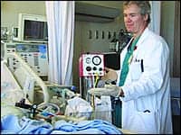
 |
Danny Sedillo, RRT, and Lisa Refrezo, CNS, of Torrance Memorial Medical Center, perform a ventilated patient simulation using capnography monitoring equipment. |
Torrance Embraces Capnography
At Torrance Memorial, capnography has allowed cardiovascular physicians and other staff to monitor patients’ carbon dioxide exhalations literally “breath by breath.” Before our widespread deployment of the technology, we relied solely on arterial blood gas analysis, which could potentially reduce a patient’s hemoglobin level with the repeated draws necessary to monitor a patient’s condition.
Broad use of capnography also has allowed the hospital’s “Code Blue” team to immediately respond to changes in patient status while it performs advanced cardiac life support CPR.
Our preliminary success has encouraged even more expanded use of capnography. We now use a portable EtCO2 device in our Rapid Response Teams (RRT) to rule in or out hypercapnia in lethargic patients. Torrance Memorial equipped its RRT staff with Oridion Microcap devices, which are portable capnography monitors that provide accurate, continuous monitoring for all patient types, whether nonintubated or intubated. The technology enables responders to an emergency to focus on patient need quickly.
“Instituting the use of end-tidal CO2 was well accepted by the respiratory care practitioners at Torrance,” said Pamela Michael, registered respiratory therapist and director of Pulmonary Services at Torrance Memorial. “Measuring end-tidal CO2 in cardiac arrest patients is helpful for confirming tracheal tube placement, assessing the effectiveness of chest compressions, and predicting likelihood of return of spontaneous circulation (ROSC).”
Broad Uptake
Capnography has become a standard, like pulse oximetry, to the Torrance Memorial respiratory department. It no longer does unnecessary arterial blood gas analysis just to measure a patient’s carbon dioxide levels.
For example, the hospital makes sure that patients using patient-controlled analgesia pumps are continually monitored using the Alaris IV pump system, a comprehensive intravenous medication safety system with built-in Oridion capnography modules. The capnography component provides the earliest warning possible of potential respiratory problems.
Respiratory care practitioners work closely with the physicians and nurses to utilize these devices during advanced cardiac life support and pediatric life support as recommended by the American Heart Association 2010 Guidelines for Cardiopulmonary Resuscitation and Emergency Cardiovascular Care.
This technology allows the team to have a consistent measuring tool to judge the quality and effectiveness of resuscitation efforts for our patients. It allows for real-time feedback on treatment efforts and gives all members of the team important information to optimize the care of our patients.
“Capnography use has been greatly helpful in verifying and monitoring patients in the hospital setting,” said Torrance Memorial respiratory therapist Marc Mercacdo. “In my experience, the application of capnography especially during ‘Code Blue’ situations has proven to be a good indicator for return of spontaneous circulation.”
Code response tends to rely on information provided by Oridion Capnostream 20 monitors, which are bedside capnography monitors that can be used in any hospital environment and for all patient types. Because these devices allow us to monitor patients continually, they provide both the information needed to assess patient conditions and alerts to call for help when adverse conditions arise.
Torrance Memorial also uses capnography with heart surgeries because it indirectly measures cardiac output and helps respiratory therapists to correctly access patient ventilatory needs.
Torrance Memorial was methodical in selecting the best technology to maximize protection for our patients in our unique environment. The technology has helped us protect patients by identifying cases of respiratory depression before patients were harmed. RT
Patrick Moore, RRT, Respiratory Therapy Clinical Educator at Torrance Memorial Medical Center, Torrance, Calif, is president of the California State Society for Respiratory Care – South Coast Region, a member of the faculty at El Camino College, and California Society Chair of Drive-for-COPD 2013. For further information, contact [email protected].