With the full implementation of the Affordable Care Act, pulse oximetry is poised to play an even bigger role in helping hospitals achieve the end goal of reducing readmissions through patient monitoring.
By Phyllis Hanlon
Healthcare facilities and medical professionals have been using pulse oximetry for more than eight decades to monitor patients’ oxygen levels. During that time, more sophisticated models with advanced capabilities have been developed. With the full implementation of the Affordable Care Act (ACA), pulse oximetry is poised to play an even bigger role in helping hospitals achieve the end goal of reducing readmissions through patient monitoring.
Depending upon setting and patient condition, one of two different pulse oximetry monitoring options might be used. Spot checking, or intermittent pulse oximetry, is used for low acuity monitoring, according to Mark VanderWerf, vice president of eHealth and OEM at Nonin Medical Inc. Usually performed every 2 to 4 hours to observe a patient’s recovery and condition, spot checking may be integrated into a multiparameter monitor that captures other data as well.
Continuous monitoring measures second-to-second oxygen levels and is routinely used to supervise all patients in the operating room and the intensive care unit (ICU), noted Joe Crackel, product manager for Nonin.
Continuous as Standard
In certain cases, continuous monitoring is standard, such as during pulmonary function testing (PFT) when patients wear a portable oxygen monitor. “It’s usually a Bluetooth with a wireless link. The patient can walk for 6 minutes without holding onto a machine. They wear it on their body,” said Crackel. The device not only measures oxygen saturation levels and how much the levels change from baseline, but also records the distance the patient covers during the 6-minute test.
Nonin created a wrist model pulse oximeter that stores data in memory. “CareFusion partnered with Nonin to take real-time data from the Bluetooth 3150 and export it to the medical record,” Crackel said, adding that hospital systems without electronic medical records systems can receive a stand-alone report that can be attached as a PDF to the medical record.
Continuous pulse oximetry is also used to monitor patients during sleep apnea studies. Clinicians believe a patient with sleep apnea or on opioids is at greater risk for deterioration at night, so some hospitals have implemented round-the-clock, or nocturnal, monitoring.
Recognizing the high risk for sentinel events, The Joint Commission (TJC) advocates for continuous pulse oximetry for patients on opioids for pain. The Anesthesia Patient Safety Foundation (APSF) and the Centers for Medicare and Medicaid Services (CMS) also support continuous pulse oximetry to prevent sentinel events and research has proven its effectiveness.
A 2010 landmark study1 published in Anesthesiology found a “significant drop” in several key clinical outcome measures when continuous monitoring is practiced, including 65% fewer rescue events, 48% fewer ICU transfers, and reduced annualized ICU time by 135 days. This joint Dartmouth-Hitchcock Medical Center/Masimo trial used the Masimo Patient SafetyNet remote monitoring and clinician notification system.
Paul Jansen, executive vice president of marketing and clinical development at Masimo, explained that implementing most advances in patient care is an expensive undertaking; however, pulse oximetry is relatively inexpensive to integrate into patient care. Dartmouth saved $1.5 million when using pulse oximetry, even when the cost of sensors and devices was factored in, he said.
In spite of the evidence, changing monitoring practices from intermittent to continuous does not occur easily. Jansen noted nursing staff, particularly in the ICU, cares for multiple, high-acuity patients, who are connected to multiple pieces of equipment. These medical devices, including pulse oximeters, emit alarms, which can become burdensome.
“When you try to implement continuous monitoring, you could have thousands of alarms. Nurse can’t accept this,” Jansen said. He reported that Masimo signal extraction (SET) technology has been proven to reduce the frequency of false alarms 5% of the time when patients are moving or have low perfusion. “If you keep the patient from having rescue events, you’ll more likely discharge the patient sooner and in better condition.”
Another modality, continuous pulse co-oximetry, uses multiple wavelengths of light emitters with different frequencies to monitor blood hemoglobin, methemoglobin, and carboxyhemoglobin levels and note any downward trends. Masimo’s Rainbow co-oximetry obtains SpO2 readings and pulse rate, provides lab measurements, and scrutinizes the patient’s breathing with an integrated acoustic transducer-equipped sensor applied to the neck.
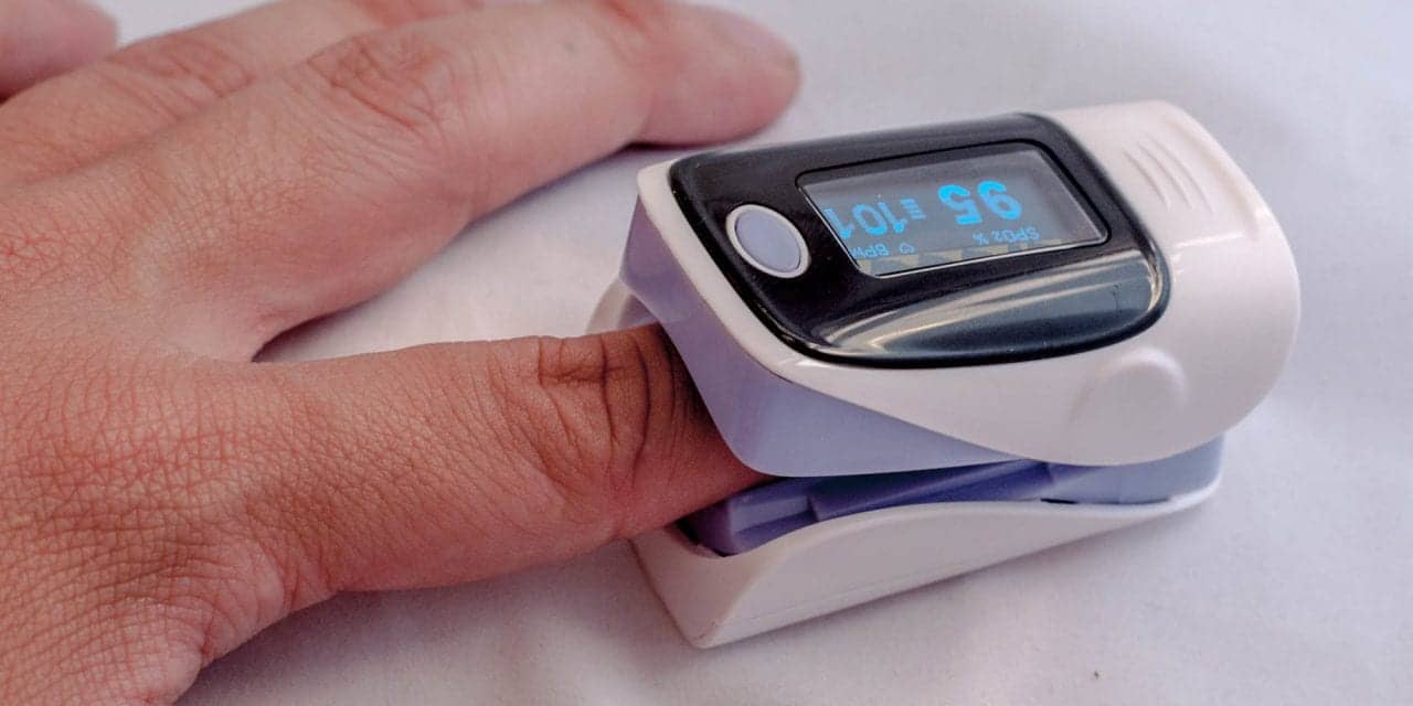
Monitoring with pulse oximetry extends beyond the hospital walls as well. Upon discharge, certain patients receive operating instructions for a remote device such as Nonin’s Bluetooth Smart Model 3230 finger pulse oximeter, winner of the 2014 Bluetooth Breakthrough Award, said VanderWerf.
The screen shows the patient how to attach the clip to the finger and the device monitors oxygen levels. If the levels fall out of recommended parameters, an alert can be sent to a call center nurse who assesses the situation. “An ‘escalation path’ protocol is put in place, based on what the device is seeing,” said VanderWerf. Monitoring via telemedicine can benefit patients with a variety of diagnoses, such as congestive heart failure (CHF), COPD, diabetes, and comorbidities.
Cerebral Oximetry Measures Tissue Oxygenation
While pulse oximetry measures hemoglobin-oxygen saturation in pulsatile arteries, cerebral oximetry, also known as regional tissue oximetry, provides a window into the adequacy of local tissue oxygenation and perfusion.
Originally used in adult and pediatric cardiac operating rooms as a noninvasive, real-time measure of the adequacy of brain oxygen delivery before, during, and after cardiopulmonary bypass, cerebral oximetry monitoring has now spread into the pediatric and neonatal intensive care units, as well as into some emergency departments, according to Henry Szymanski, RRT, senior product manager, INVOS, Covidien.
“Cerebral oximetry is useful in any acute care environment in which a patient’s brain could be exposed to regional hypo-perfusion, such as in pediatric and neonatal intensive care areas and postoperative cardiac surgery,” Szymanski said.
Regional oximetry takes a continuous, noninvasive, and site-specific venous measurement in the tissue capillary bed, which represents the amount of oxygen remaining after tissue has extracted what is needed to meet metabolic demand.
“Many of the physiologic measures used by clinicians are global ‘supply-side’ data, such as peripheral oxygen saturation, hemoglobin, mean arterial pressure, etc. These variables are reflective of flow and content going to the organs and do not indicate whether the flow and content are adequate,” Szymanski said. “Only assessment of the venous side of the metabolic equation can provide an indication of whether these upstream supply-side variables are adequate.”
Covidien’s INVOS 5100C cerebral/somatic oximeter uses near infrared light spectroscopy (NIRS) to determine the fraction of hemoglobin-oxygen saturation in tissue under a sensor, which can be applied anywhere on the body. The INVOS system isolates a measurement attained through spatial resolution from deep tissue that aids the clinician in determining whether the oxygen supply to the brain meets the oxygen demand, a process called “assessing adequacy of perfusion.”
Szymanski said, “Spatial resolution occurs when the depth of penetration of the near infrared light is reflected back to the detectors, and is proportional to the distance between the light source and the detector.” A shallow detector is spaced 3 cm from the light source and measures shallow tissue, while a deep detector is spaced 4 cm from the light source, and this measurement represents both shallow and deep tissues.
When the shallow measurement is subtracted from the deep measurement, the resulting value is that of the deep tissue. “The depth of the measurement made by the INVOS monitor is approximately 2-3 cm, a depth adequate to penetrate the extra-cranial tissue and to provide a highly reliable measure of brain tissue oxygenation in adult and pediatric patients,” Szymanski added.
Reducing Postoperative Events
Providing an indication of cerebral perfusion adequacy is particularly important during cardiac surgery where hemodynamic perturbations can be severe. Prolonged cerebral desaturation events commonly occur during cardiac surgery and have been associated with an increase in postoperative complications, including neurologic injury, extended mechanical ventilation, and prolonged stays in the ICU.
“Minimizing complications, especially neurological, could conceivably reduce postoperative time in the ICU and possibly streamline the transition of the patient to the general care floor and discharge,” Szymanski said. “While this concept has yet to be studied, there is evidence that shows lower baseline preoperative cerebral saturation is associated with poorer outcome in adult cardiac surgery patients. This evidence may help support the concept and study of preoperative screening as a means to identify high-risk patients before they enter the surgical stream of care.”
Brian Kane, product manager for Nonin, pointed out that cerebral oximetry can be used effectively to help guide cardiopulmonary resuscitation. “For instance, if a cannula is out of position, blood is not reaching the brain. This would not have been discovered in the past. You have to use cerebral oximetry to know this,” he said. “Also, cerebral oximetry is proven clinically to have the ability to decrease the incidence of postoperative cognitive dysfunction and reduce the incidence of stroke. It’s a fast and upcoming medical application.”
Moreover, cerebral oximetry can evaluate whether oxygen is reaching its ultimate destination, such as target tissue, even in extreme circumstances. “It can detect the level of oxygen, even in people who are so poorly perfused, they have no pulse,” Kane said.
During the last few years, the healthcare industry has been adopting interoperability in an effort to create a more efficient and effective patient care environment. Kane noted that Nonin Medical’s EQUANOX Model 7600 Regional Oximetry System connects to Philips IntelliVue Monitor via IntelliBridge Open-Interface Model.
The INVOS monitor can interface with many electronic medical record systems and other clinical data recording systems and is also compatible with some common middleware or gateway systems, such as Capsule, which provides access to a variety of EMR systems, said Szymanski.
Learning Curve
Becoming comfortable with any new device requires a learning curve that depends on the clinician’s experience and the complexity of the device. Pulse oximetry is relatively straightforward; the clinician applies a sensor to the finger or the neck. Sensor placement on pulse CO-oximetry requires more precision than standard pulse oximetry, since the emitter and detector have to line up.
Cerebral oximetry demands a certain level of expertise, although the learning curve is minimal. Kane explained that in pediatric patients, the lead is placed over the renal bed to find perfusion status. For adults, clinicians place two patches on the patient’s head, one on each side of the forehead, which measure the two hemispheres of the brain and display the data on a screen.
“Manipulations can have an immediate impact on the oxygen saturations. For example, the patient on a bypass with low saturation needs to have the pump flow or CO2 increased. When you do this, you will see immediate changes,” Kane said.
Szymanski said, “Another somatic site includes positioning the sensor over the calf muscles to detect potential compromise to lower extremity perfusion state.” Clinicians are also using INVOS to monitor challenging patients, such as those in shock, with acute respiratory distress syndrome (ARDS) or undergoing extracorporeal membrane oxygenation (ECMO).
Using a cerebral oximetry monitor and integrating it into clinical practice is a matter of understanding what the measurement represents, according to Szymanski. “Typically, the learning curve is the clinician becoming comfortable utilizing the monitor and recognizing that the measurements represent important physiologic status and then incorporating the information during patient assessment and clinical decision-making,” he said.
Furthermore, most manufacturers offer some type of educational support post-purchase to help acclimate users to the device. Covidien provides in-person and online training through the Professional Affairs and Clinical Education website and through peer-to-peer education programs with clinicians who have in-depth experience using INVOS monitors. Nonin provides a number of training resources and product support, including “real-person” customer care, technical service, videos, sensor application wall charts, and operators’ manuals.
Although noninvasive monitoring technologies, such as cerebral oximetry, are basically safe, clinicians must understand any limitations associated with use of the technology. “The range of normal values for cerebral and somatic venous oxygen saturation is wide, and low values do not necessarily indicate a ‘bad’ condition. Proper use of the INVOS monitor usually involves establishing a patient’s baseline and then assessing the impact of changes from baseline,” said Szymanski.
Cerebral oximetry is not intended to replace standard monitoring parameters; rather, it is designed to provide clinicians with additional tissue-based information and insight into changes in tissue oxygen demand and whether that demand is being satisfied.
RT
Phyllis Hanlon is a contributing writer to RT. For further information, contact [email protected].