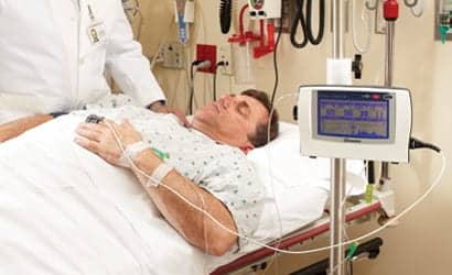
Capnography and capnometry are valuable tools for many applications when used in conjunction with other clinical data.
By Roy Ramirez, RRT-NPS, RPFT; Terri Pickar, CRT-NPS; Sam Frens, RRT-NPS; James Cappon, MD; and John Cleary, MD
Potentially preventable patient deaths have occurred because of ventilation-related problems. Life-threatening airway malfunctions can be averted with the use of continuous capnometry monitoring. Changes in circulatory and respiratory status can be detected sooner with capnography than with pulse oximetry.
Capnometry: Current Technologies
Capnometry (digital display of data) is the measurement of fractional carbon dioxide (Fco2) tidal gas at the airway opening.1,2 Capnography is the graphic display of measured Fco2 and can be time or volume based. Time-based capnography is most widely known as end-tidal carbon dioxide (PETCO2) monitoring. Volume-based capnography is known as volumetric CO2 monitoring (Vco2); it uses a combined CO2 sensor and a pneumotachometer. This allows for a calculation of the net volume of CO2 expired (Vco2) by the patient expressed as a volume of gas in mL/min rather than a partial pressure or gas fraction.
Vco2 is affected by ventilation, pulmonary circulation/perfusion, and, to a lesser degree, diffusion. It is a valuable marker in assessing the cardiorespiratory status of the ventilated patient. The Vco2 provides information about CO2 elimination, alveolar ventilation (Valv) and physiologic dead space (VDphy). All three parameters provide important data about the physiologic state of the patient. This combined information helps us to optimize ventilator support and understand the physiologic status of the patient.

Volumetric Capnography
The volumetric capnogram is comprised of three separate phases (Figure 1).
Phase 1 represents volume of gas exhaled from the upper airways anatomic dead space (VDanat).
Phase 2 is the transitional phase from upper to lower airway ventilation, and it typically depicts changes in pulmonary perfusion. For example, a decrease in the slope in phase 2 indicates a decrease in perfusion. This slope change is due to an increase in alveolar dead space that occurs in ventilated alveolar units with decreased or absent perfusion.
Phase 3 reflects alveolar gas exchange and represents changes in gas distribution. An increase in the slope of this phase indicates maldistribution of gas delivery. This might be seen in an asthmatic patient.
Capnography has been erroneously judged as unreliable for continuous CO2 monitoring because of the variability between Paco2 measurements and PETCO2 readings. The information obtained from capnography needs to be evaluated carefully in conjunction with other clinical data and a thorough understanding of physiology and gas exchange. Factors that affect the Paco2 and PETCO2 gradient (P(a-ETCO2) can include air leaks, changes in airway physiology, and alveolar dead space, as well as alterations in cardiac output caused by such conditions as shock, cardiac dysfunction, or increased pulmonary vascular resistance. In these conditions, there is a decrease in Vco2 as an indicator of ventilation/perfusion ratio (V/Q) mismatching secondary to changes in either anatomic or physiologic dead space. The normal P(a-ETCO2) gradient is 4 to 5 mm Hg, which represents normal dead-space ventilation (VD/VT).

The P(a-ETCO2) is directly proportional to the alveolar dead space. Thus, as alveolar dead space increases, so does the P(a-ETCO2) gradient. In the care of our patients, when a blood gas is obtained, the P(a-ETCO2) gradient is observed, the Vco2 trended, and any physiologic changes occurring in the patient are noted. Observation of these three factors enhances our ability to interpret and correctly apply this data.
In the clinical setting, stable alveolar ventilation and CO2 production and changes in Vco2 can be attributed to a lung perfusion factor (Figure 2). This needs to be considered when ventilator changes are made to improve gas exchange in a patient. If the post-manipulation blood gas reflects no change in Paco2, a perfusion problem should be considered.3 Without a device such as the NICO cardiopulmonary management system (Philips-Respironics, High Acuity-Hospital Respiratory Care, Carlsbad, Calif) providing simultaneous Vco2 data, this consideration would be unavailable, and clinicians would be compelled to continue to adjust the ventilator to lower the Paco2, and possibly further compromise pulmonary perfusion.
Sampling Devices
There are two exhaled gas sampling methods: sidestream and mainstream sampling. A mainstream capnometer has an airway adaptor cuvette that is attached in-line, close to the artificial airway. The cuvette contains an infrared light source and sensor that detects CO2 in the exhaled gas. The sidestream capnometer uses a sample line that attaches to an adaptor at the airway opening. It pulls gas continually into a chamber for analysis. This method requires a lightweight adaptor to be added to the breathing circuit.
Airway Management
Capnography can be an essential tool for the determination of endotracheal tube (ETT) placement. In the neonatal population, capnography can more quickly and accurately determine tracheal intubation than can other clinical assessments.4
The American Heart Association (AHA) strongly supports the use of capnography to confirm tracheal intubation in infants and children with perfusing rhythms. To confirm ETT position, the AHA recommends using colorimetry or capnography to detect exhaled CO2. In addition, the American Association for Respiratory Care has published guidelines for the use of capnography during mechanical ventilation.5
When repositioning the patient post intubation, an ETCO2 device will quickly alert the clinician to a dislodged or obstructed ETT. Furthermore, any problems with an ETT that is obstructed or partially kinked would immediately be reflected by a change in the PETCO2.6
Optimizing Mechanical Ventilation
Mechanical ventilation is not without risks.7 Minimizing the duration of mechanical ventilation is very important in the critically ill patient. The shorter the duration of ventilatory support, the less exposed a patient will be to its adverse effects, such as ventilator induced lung injury (VILI), airway injury, nosocomial pneumonia, and excessive use of pharmacologic sedation, all potentially predisposing to increases in morbidity and mortality.8
Continuous capnography in the ventilated patient offers several benefits, including optimizing mechanical ventilator support, evaluating pulmonary capillary blood flow and cardiac output, and weaning to extubation.
Tidal Volume
During positive pressure ventilation, the quantity of CO2 expired is controlled by adjusting the total minute ventilation (= VT times respiratory rate). In lung-protective ventilation strategies, accurate VT assessment is essential. The volume displayed by the ventilator as delivered may not reflect actual volume of gas delivered to the lungs.9 When measuring VT with a pneumotachometer between the ventilator circuit and the endotracheal tube, the ventilator circuit compliance and other circuit variables are no longer able to add false data, and an accurate delivered VT is displayed.
Alveolar Minute Ventilation
The minute ventilation value is the calculated volume of total measured rate x exhaled VT. It does not reflect the amount of gas participating in gas exchange. Volumetric capnography provides us with measured alveolar minute ventilation (MValv). This is the volume of gas that participates in gas exchange at the alveolar level.10 With the ability to monitor MValv, the clinician is able to observe changes in ventilation when there are compliance fluctuations.

For example, in a patient who develops a distended abdomen on pressure control ventilation, the MValv would decrease as the abdomen distention progressed.
The clinician can now observe if increasing ventilator positive pressure has a positive effect (higher MValv) or a negative effect (ETT air leak), at which time the respiratory rate could be increased, thereby effectively increasing MValv.
PEEP Management
Achieving the appropriate positive end-expiratory pressure (PEEP) level is important in managing the mechanically ventilated patient with acute lung injury. The goal is to deliver the appropriate PEEP to achieve the highest oxygenation with the lowest Fio2, the highest pulmonary compliance, and the optimal cardiac output. One method of titrating PEEP when using a capnograph is to monitor Vco2. When we have increased PEEP while monitoring Vco2 clinically, we have observed a transient decrease in Vco2, usually a <20 % change. When PEEP support exceeds patient tolerance, the Vco2 reflects a decrease of >20%, which is largely due to a decrease in pulmonary perfusion secondary to a decrease in systemic venous return. Decreased pulmonary blood flow reduces CO2 (Vco2) transport from the tissues to the pulmonary vasculature, which reduces CO2 elimination.11
Weaning
Weaning ventilated patients often requires multiple blood-gas analyses, but using this method provides only a single point of information. It can also lead to anemia in premature infants.12 Capnography provides information on breathing patterns and illustrates the importance of breathing consistency before successful weaning can occur.13 In the event that a patient deteriorates during a ventilator weaning trial, it can be identified earlier, and previous ventilator settings can quickly be restored.
A clinical study of 45 infants13 reported the use of capnogram parameters to predict the success of extubation in infant and children. It was demonstrated that the ratio of physiologic dead space to tidal volume (VD/VT) <0.5 predicted a successful extubation. Conversely, a VD/VT >0.65 identifies a patient at high risk for post-extubation.
Neonatal Applications
Measurement of neonatal pulmonary function was first performed in the 1950s. This initial work helped to define the groundwork for neonatal pulmonary pathophysiology,14-17 but progress in the study of capnography in the neonate was not possible until the development of mainstream airway cuvettes.
The development of the new and improved mainstream capnographs that were lightweight and had a dead space of less than 1 mL opened the door to utilizing capnography in the NICU. These cuvettes reliably provide measurements in preterm infants.18,19 In spite of these ground-breaking developments, capnography has not been embraced by clinicians to monitor alveolar gas exchange in the neonate. Some of the limitations include technical problems such as sensor dead space and short expiratory time in neonates with stiff lungs20,21 and capnograph resolution in the presence of high respiratory rates. These issues are exacerbated in very low birth weight (VLBW) infants of <1,500 grams.22
In the neonate, analysis of the single breath carbon dioxide waveform requires comparison with the typical pattern as illustrated in Figure 1. Neonatal phase 1 should be short. Phase 2 should rise with a steep upward slope. Phase 3 is usually absent in neonates; if it is present, it is often small with slope a function of lung growth.23 Interpreting phase 3 must be done with care, but the presence of phase 3 is an indication that alveolar gas was sampled.
In infants with normal pulmonary function and normal ventilation-to-perfusion ratio (V/Q), the PETCO2 can be a good estimation of the Paco2.24 In critically ill patients, the V/Q ratio is often abnormal with a resulting increase in the P(a-ETCO2) gradient.
Although much progress has been done since the early use of capnography in the neonate, we still face technical limitations in measuring CO2 signals in VLBW infants. In addition, much needs to be learned about how small and immature lungs physiologically differ and the resultant impact on gas exchange, particularly phase 2 and phase 3.

Congenital Heart Disease
Congenital heart diseases can be categorized into two broad groups: cyanotic and noncyanotic. Cyanotic lesions are defects that result in low blood oxygen levels. Cyanosis results in an abnormal blood flow from the right side of the heart to the left side of the heart, bypassing the lungs and creating a right-to-left shunt. Some congenital heart defects that cause cyanosis include: tetralogy of Fallot, transposition of the great vessels, tricuspid atresia, pulmonary atresia, and hypoplastic left heart.25
Noncyanotic congenital heart disease includes lesions in which blood returning to the right side of the heart passes normally through the pulmonary vasculature. The common forms of noncyanotic congenital heart defects are conditions where there is an opening in one of the walls separating the heart chambers or obstruction to a cardiac valve. Some types of noncyanotic heart disease are: coarctation of the aorta, atrial septal defect, ventricular septal defect, atrioventricular septal defect, patent ductus arteriosus, and aortic stenosis.25
Any factor that alters cardiovascular function can affect the transport of CO2 to the lungs. In the absence of changes in the respiratory status of the patient, it should alert clinicians to possible changes in the cardiovascular function of the patient. In patients with cyanotic heart disease, the alveolar ventilation is usually normal. Pulmonary perfusion may be abnormal, however, and that can result in a V/Q mismatch. Therefore, PETCO2 cannot be used as a reliable estimate of Paco2 in the congenital heart disease patient.
Capnography usage is particularly helpful in monitoring pulmonary perfusion in the postoperative cardiac patient with either a Blalock-Taussig or central shunt (Figure 4). These palliative conduits from the systemic arterial circulation to the pulmonary artery are placed to provide reliable pulmonary blood flow in some forms of cyanotic heart disease, in which pulmonary blood flow is much decreased or absent. In the potentially life-threatening event of shunt clotting, there would be a significant decrease in pulmonary blood flow, with a subsequent decrease or absence of EtCO2.
Conclusion
At our facility, we have progressively embraced capnography as part of our patient care process. We provide frequent education for staff to ensure they are aware of the many applications of this technology. Once the decision has been made that a patient requires endotracheal intubation, our standard of practice is to use a colorimetric CO2 detector to verify ETT placement. The proper placement of the tube is further clinically confirmed by auscultation for breath sounds bilaterally and is followed with an immediate chest x-ray. We then place the patient on a capnography monitor and use the data in our treatment of the patient. Capnography is the standard of care for all cardiac, near term infants and critically ill pediatric patients on conventional ventilation. We also use it to evaluate readiness for extubation. It is our belief that a comprehensive patient-management approach utilizing capnography provides optimal respiratory care and patient safety support and improves outcomes in the mechanically ventilated child.
RT
Roy Ramirez, RRT-NPS, RPFT, is clinical educator, respiratory therapy services and ECLS coordinator; Terri Pickar, CRT-NPS, is shift lead; Sam Frens, RRT-NPS, is shift lead; James Cappon, MD, is medical director, quality and patient safety; and John Cleary, MD, is associate director, cardiovascular intensive care and ECLS director, neonatology, Children’s Hospital of Orange County, Orange, Calif. For further information, contact [email protected].
References
- Breen PH. Carbon dioxide kinetics during anesthesia: pathophysiology and monitoring. In: Respiration in Anesthesia: Pathophysiology and Clinical Update. Philadelphia: WB Sounders. Anesthesiology Clinics of North America. 1998;16:259-293.
- Dang K, Breen PH. Ambulatory capnography. RT. 1998;11(4):25-30.
- Burrows FA. Physiologic dead space, venous admixture, and the arterial to end-tidal carbon dioxide difference in infants and children undergoing cardiac surgery. Anesthesiology. 1989;70:219-25.
- Roberts WA, Maniscalco WM, Cohen AR, Litmans RS, Chhibber A. The use of capnography for recognition of esophageal intubation in the neonatal intensive care unit. Pediatr Pulmonol. 1995;19:262-8.
- McArthur DC, AARC. AARC Clinical Practice Guideline. Capnography/Capnometry during mechanical ventilation—2003 revision and update. Respir Care. 2003;48:534-9.
- Palmon S, Maywin L, Moore L, Kirsch J. Capnography facilitates tight control of ventilation during transport. Crit Care Med. 1996:24:608-11.
- Mead J, Takishima T, Leith D. Stress distribution in lungs: a model of pulmonary elasticity. J Appl Physiol. 1970;28:596-608.
- Orlowski JP, Ellis NG, Amin NP, Crumine RS. Complications of airway intrusion in 100 consecutive cases in a pediatric ICU. Crit Care Med. 1980;8:324-31.
- Cannon ML, Cornell J, Tripp-Hamel DS, et al. Tidal volumes for ventilated infants should be determined with a pneumotachometer placed at the endotracheal tube. Am J Respir Crit Care Med. 2000;162:2109-12.
- Laurence M. The essentials for patient care and evaluation. In: Pulmonary Physiology in Clinical Practice: The Essentials for Patient Care and Evaluation. St Louis: C.V. Mosby; 1987.
- Johnson JL, Breen PH. How does positive end-expiratory pressure decrease pulmonary CO2 elimination in anesthetized patients? Respir Physiol. 1999;118:227-36.
- Obladen M, Maier RF. Recombinant erythropoietin for prevention of anemia in preterm infants. J Perinat Med. 1995;23:119-26.
- Hubble CL, Gentile MA, Tripp DS, Craig DM, Meliones JN, Cheifetz IM. Deadspace to tidal volume ratio predicts successful extubation in infants and children. Crit Care Med. 2000;28:2034-40.
- Gerhardt TO, Banclar E. Measurement and monitoring of pulmonary function. Clin Perinatol. 1991;18:581-609.
- Karlberg P, Cook CD, O’Brien D, Cherry RB, Smith CA. Studies of respiratory physiology in newborn infants. II. Observations during and after respiratory distress. Acta Paediatr. 1954;43:387-411.
- Nelson NM, Prod’hom LS, Cherry RB, Lipsitz PJ, Smith CA. Pulmonary function in the newborn infant: 1. Methods: ventilation and gaseous metabolism. Pediatrics. 1962;30:963-74
- Cook CD, Cherry RB, O’Brien D, Karlberg P, Smith CA. Studies of respiratory physiology in the newborn infant. Observations in normal and full-term infants. F Clin Invest. 1955;34:975-82.
- Meredith KS, Monaco FJ. Evaluation of a mainstream capnometer and end-tidal carbon dioxide monitoring in mechanically ventilated infants. Pediatr Pulmonol. 1990;9:254-9.
- Arsowa S, Schmalisch G, Wauer RR. Simultaneous measurements of end-expiratory and transcutaneous carbon dioxide partial pressure in ventilated premature and newborn infants. Klin Padiatr. 1977;209:47-53.
- Proquitté H, Krause S, Rudiger M, Wauer RR, Schmalisch G. Current limitations of volumetric capnography in surfactant-depleted small lungs. Pediatr Crit Care Med. 2004;5:75-80.
- Kirpalani H, Kechagias S, Lerman J. Technical and clinical aspects of capnography in neonates. F Med Eng Technol. 1991;15:154-61.
- Tirosh E, Bilker A, Bader D, Cohen A. Capnography in spontaneously breathing preterm and term infants. Clin Physiol. 2001;21:150-4.
- Ream RS, Schreiner MS, Neff JD, et al. Volumetric capnography in children. Influence of growth on the alveolar plateau slope. Anesthesiology. 1995;82:64-73
- McDonald JJ, Montgomery VL, Cerrito P, Parrish CJ, Boland KA, Sullivan JE. Comparison of end-tidal CO2 and PaCO2 in children receiving mechanical ventilation. Pediatr Crit Care Med. 2002;3:244-9.
- Heart Center Encyclopedia. 2006. Hypoplastic left heart syndrome. Available at: www.cincinnatichildrens.org/health/heart-encyclopedia/anomalies/hlhs.htm. Accessed November 26, 2008.