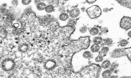
DLCO testing is useful in assessing lung function and lung health in infected and recovering COVID-19 patients.
By Bill Pruitt, MBA, RRT, CPFT, AE-C, FAARC
Research on recovering COVID-19 patients has shown a considerable prevalence of altered diffusing capacity of the lung for carbon monoxide (DLCO) percentage in survivors. Carbon monoxide (CO) is used to test the transfer of gases from the alveoli into the bloodstream. It behaves like oxygen in this transfer from lung to blood as it diffuses through the lung/vascular tissues and also, like oxygen, binds to hemoglobin once it has crossed the alveolar-capillary (a/c) membrane. A normal versus reduced DLCO can help distinguish between different lung diseases and pathologies. This article will explore the use of DLCO testing in assessing lung function and lung health in infected and recovering COVID-19 patients.
DLCO Testing
The most common DLCO test involves the patient breathing in a closed circuit. At the start of the test, the patient is instructed to exhale fully to residual volume (RV) then quickly inhale fully to total lung capacity (TLC). At the beginning of inhalation, a valve opens to provide a mixture of gases: 0.3% CO, 0.3% tracer gas, 21% oxygen, and balance nitrogen. (The tracer gas is an inert gas, usually helium, methane, or neon.) Next, the patient has a 10-second breath hold at TLC (allowing the transfer of gases to occur at the a-c membrane) followed by a full exhalation.
An analysis of a sample of the exhaled gas is performed by the PFT machine after discarding the initial exhaled gas to exclude the gas in the deadspace. The difference between the initial gas and exhaled gas provides a measurement of the diffusion in CO and difference in the inhaled/exhaled tracer gas (which does not diffuse) provides a measure of alveolar volume. If a recently measured hemoglobin is known it is included for the calculation, otherwise the test will assume a normal Hg and COHg to complete the test results. Anemia can reduce the DLCO since there is less Hg available for binding the CO and correcting results for anemia will result in an increase in the reported DLCO (reported as DLCO adjusted). An average of two or more measurements is used to calculate the DLCO in this procedure (called a single breath DLCO test) and the units of measure are cc CO/sec/mmHg.1
The measurement of gas exchange in the lung by using CO is affected by:
- diffusion coefficient of the gas used in testing
- surface area of the a-c membrane
- thickness of the a-c membrane
- blood volume in the pulmonary capillaries
- distribution of inspired gas
- hemoglobin (results are adjusted for anemia)
Pulmonary and cardiovascular problems are reflected in the last five of these six factors. Damaged or destroyed alveoli (as in emphysema) can reduce the surface area of the a-c membrane. Fibrosis can cause a thickening of the a-c membrane. Blood volume in the pulmonary capillaries is decreased by formation of clots or vasoconstriction leading to increased deadspace ventilation. Distribution of inspired gas can be altered by mucus plugs and bronchoconstriction leading to increased shunt. Anemia decreases hemoglobin and reduces the amount of CO that moves across the a-c membrane.
Another factor that can reduce diffusion capacity occurs with carbon monoxide poisoning (or excessive exposure to CO) which blunts the ability of hemoglobin to bind with the test gas containing carbon monoxide.
Severe Acute Respiratory Syndrome (SARS)
SARS is caused by the coronavirus and first became a high priority outbreak in humans with the Guangdong, China outbreak in 2002. Referred to as SARS-CoV, this infection has had 8,096 confirmed cases with 744 deaths (10.8% mortality). The next outbreak of the coronavirus in humans occurred in 2012 in Saudi Arabia (referred to as Middle East Respiratory Syndrome or MERS-CoV) with 2,519 cases and 866 deaths (34.4% mortality).2 The world-wide pandemic of SARS-CoV-2 (also referred to as COVID-19) first appeared in late 2019 in Wuhan, China. According to the latest figures from the World Health Organization, there have been 245,373,039 cases and 4,979,421 deaths (2.0% mortality).3 The three strains of coronavirus have many common points in the clinical presentation including fever, fatigue, non-productive cough, myalgia, dyspnea, sore throat, diarrhea, nausea and/or vomiting, dizziness, and headache. There are overlapping features in all three of these coronaviruses as well as some discrete features from one to the next. Around 80% of the COVID-19 cases have mild disease, 15% have severe disease, and some 5% have critical disease.4
DLCO in SARS infection
One feature that the three coronaviruses have in common is a reduced diffusion capacity (measured by DLCO) in patients who have survived and are in the post-acute phase of the disease. (Note: this is also a common feature of H1N1 influenza survivors and ARDS survivors). Some COVID-19 and ARDS survivors also show signs of restrictive disease at 3 months and 6 months after recovery.5 The level of reduction in DLCO is linked the severity of the disease. Research in COVID-19 divides patients into mild, moderate, and severe categories with severe cases often defined by having any of the following characteristics: shortness of breath, respiratory rate >30 breaths/min, SpO2 <93% at rest on room air, PaO2/FiO2 <300 mmHg, respiratory failure requiring mechanical ventilation.6
In a study6 published in 2020 examining survivors of COVID-19 at 30 days post-discharge from the hospital, a total of 57 patients were examined; 40 of these were classified as non-severe and 17 were classified as severe. Of the 57 enrolled, 30 were found to have an abnormal DLCO, defined as being less than 80% of predicted value. When comparing non-severe to severe patients, 17 of the 40 non-severe cases (42.5%) had a DLCO <80% of predicted while in the severe cases, 13 of the 17 (76.5%) had a DLCO <80% of predicted.6
Another study examined 119 survivors in a one year follow-up and divided the survivors into 3 categories; those who were asymptomatic (n=9), non-severe (n=82), and severe (n=28). Severe cases were defined as having fever or suspected respiratory infection, plus one of the following: respiratory rate >30 breaths/min, severe respiratory distress, or SpO2 ≤93% on room air. Of the 119 patients, 47 had an abnormal DLCO (<80% of predicted value). In the breakdown per category, one patient (11%) was from the asymptomatic group, 31 (38%) from the non-severe group, and 15 (54%) from the severe group.7 The researchers assessed other pulmonary function measures and stated, “The gradual decline in lung diffusion capacity among survivors was consistent with varying degrees of severity. There was no significant difference in other measures (TLC, RV/TLC, FVC, FEV1, FEV1/FVC, and MMEF 75/25) among COVID-19 survivors with different disease severity.” They went on to summarize their findings to say, “Lung damage might be related to pulmonary fibrosis. Further long-term research is needed to understand the mechanisms underlying long-term SARS-CoV-2-related pulmonary function damage.”7
Some patients with mild COVID-19 infection will have a decrease in DLCO without showing evidence of airflow restriction with spirometry. These patients may or may not be hospitalized. Data suggests that some two-thirds of these cases will return to their previous health status within weeks after recovery from the infection, but one-third will continue to exhibit symptoms.4 From the observation of trends it is estimated that some 30-60% of COVID-19 patients will have enduring symptoms following resolution of the acute disease. Fatigue and dyspnea are common in these cases which are being described as “long COVID” or post-COVID syndrome.8 Reduced diffusion is likely a contributing factor in fatigue and dyspnea.
Possible Causes of Reduced DLCO
As discussed earlier, there are a number of possible causes for the decrease in DLCO for patients who survived COVID-19 infection. Autopsies have shown destruction of alveolar structures, which decrease the surface area of the a-c membrane, and interstitial fibrosis, which thickens the a-c membrane. Some patients may have abnormalities in the capillary bed including formation of microthrombus that stops blood supply to alveoli (reducing perfusion and increasing deadspace ventilation).
Finally, some patients with severe COVID-19 infection have had mucous plugs in the small airways that alter the distribution of alveolar ventilation and increase shunt.6
Improved Lung Function Over Time
According to an April 2021 study, abnormalities in PFT can improve over time.9 Enrolled patients (n=145) had PFTs done 60 and 100 days post-COVID-19 onset. This was prospective, multicenter, observational trial that, so far, has not had other similar trials in looking at periodic follow-up on lung function.9 A sample of some of the pertinent PFT results are shown in Table 1.
| TEST PERFORMED | 60-day visit | 100-day visit | Improvement |
| Spirometry | 27% had restrictive pattern | 22% had restrictive pattern | +26.1% |
| Body plethysmography | 11% had reduced TLC | 11% had reduced TLC | No change |
| DLCO < 80% predicted | 31% had reduced DLCO | 21% had reduced DLCO | +30.3% |
The authors commented that there was a moderate but significant improvement in most of the PFT measurements over the time from 60 to 100 days post onset of the disease. They also noted that lung function at the time of discharge, “revealed a high rate of abnormalities indicative of potential interstitial lung disease.” 9
Conclusion
As stated in one of the articles regarding this topic, “The long-term impact that SARS-CoV-2 infection has on PFT will continue to emerge over the coming years.” The article also stated, “There is however no data yet on longer-term follow-up or with respect to the impact of targeted interventions, for example, use of systemic steroid treatment.”
Finally, “there also needs to be attention directed to the underlying cause of the reduction in diffusing capacity observed, and the impact the disease has on the pulmonary vasculature requires investigation.”8 Reduced DLCO levels are “thought to be caused by a combination of changes in the lung parenchyma combined with a variety of pathophysiologies in the pulmonary vasculature.”4 DLCO is an important measure to assess patients who survive COVID-19 early in the recovery process with subsequent tests to follow improvement. Rehabilitation efforts can be monitored and refined using DLCO where spirometry and other lung function tests may not show abnormalities and/or improvements.
RT
Bill Pruitt, MBA, RRT, CPFT, AE-C, FAARC, is a writer, lecturer, and consultant and recently retired from over 20 years teaching at the University of South Alabama in Cardiorespiratory Care. He also volunteers at the Pulmonary Clinic at Victory Health Partners in Mobile, AL. For more information, contact [email protected].
References
- Modi P, Cascella M. Diffusing Capacity Of The Lungs For Carbon Monoxide. [Updated 2021 Mar 24]. In: StatPearls [Internet]. Treasure Island (FL): StatPearls Publishing; 2021 Jan-. Available from: https://www.ncbi.nlm.nih.gov/books/NBK556149/.
- Rabaan AA, et al. SARS-CoV-2, SARS-CoV, and MERS-COV: a comparative overview. Infez Med. 2020 Jun 1;28(2):174-84.
- World Health Organization website on COVID-19. https://covid19.who.int/. Accessed Oct. 2021.
- From ndd White Paper “Relevance of DLCO and lung function testing for optimized patient management in Long-COVID “
- Nalbandian A, et al. Post-acute COVID-19 syndrome. Nature medicine. 2021 Apr;27(4):601-15.
- Huang Y, Tan C, Wu J, Chen M, Wang Z, et al. Impact of coronavirus disease 2019 on pulmonary function in early convalescence phase. Respiratory research. 2020 Dec;21(1):1-0.
- Yan X, et al. Follow-up study of pulmonary function among COVID-19 survivors 1 year after recovery. The Journal of Infection. 2021 May 29.
- Thomas M, et al. Pulmonary function and COVID-19. Current opinion in physiology. 2021 Mar 26. 21:29-35
- Sonnweber T, et al. Cardiopulmonary recovery after COVID-19: an observational prospective multicentre trial. European Respiratory Journal. 2021 Apr 1;57(4).

Respiratory Simulators from Hans Rudolph Inc
Hans Rudolph Inc, manufacturers of respiratory products since 1938, offers two lung simulators.
The DLCO Simulator with EasyLab QC Software is used by pulmonary labs worldwide for many years. This patented device and software allow the quality control and testing of respiratory devices that measure the diffusion lung capacity of patients on a daily basis and during drug delivery trials.
COVID has emphasized the importance of daily quality control of the DLCO devices being used in hospitals worldwide.
Also available from Hans Rudolph is a Flow/Volume Simulator for research and development, and testing and calibrations on aerosol devices, peak flow meters, spirometers, and other devices requiring the generation of the ATS and custom breathing waveforms.
Hans Rudolph Inc, Makers of Respiratory Valve since 1938
800-456-6695 or 913-422-7788
[email protected]
www.rudolphkc.com


