RCPs must have experience handling patients with neuromuscular diseases and administering therapeutic interventions to successfully treat patients with ALS.
Amyotrophic lateral sclerosis (ALS) is a progressive neuromuscular condition characterized by muscular weakness, wasting, fasciculation, and increased reflexes.1 It is more commonly known as Lou Gehrig’s disease for the baseball player who succumbed to this condition in 1941.
ALS is the most common degenerative motor disease in adults.2 It is a relentlessly progressive disorder that involves both the lower motor neurons in the brain stem and spinal cord and the upper motor neurons in the motor cortex. This coexistence of both upper and lower motor neuron findings is the distinctive feature of ALS. The confirmation of ALS is usually based on a lengthy elimination of differential diagnoses, and electromyography is typically the definitive test. It provides an objective examination of the lower motor neurons, and, in many cases, may document more widespread neurogenic changes than are evident in the findings of the clinical examination. Other diagnostic tests employed may include MRI studies, routine laboratory analyses, sural-nerve biopsies, muscle biopsies, and bone-marrow biopsies.2
In general, ALS is classified as sporadic, with an annual incidence of about one to two per 100,000 people, and it is slightly more common in men.2 Approximately 30,000 US residents currently have the disease, and it is most often diagnosed in middle age.1 Although there are rare reports of ALS onset in people 13 to 29 years old, the incidence of ALS peaks among those 55 to 75 years old. It is thought that the onset of ALS in patients older than 75 may be underreported due to the fact that many of the symptoms may be attributed to the debility of old age or to other medical conditions.3,4 Given the similar incidence reported from around the world, the occurrence of ALS seems to be independent of socioeconomic, racial, or geographic differences.5 There has been a modest increase in the reported incidence of ALS in several countries in the past 20 to 30 years; some researchers theorize that the increase is due to unknown environmental or exogenous factors. A few geographic clusters of patients with ALS have also been reported, but their significance is not clear.6 Some researchers believe that the reported increases in incidence may simply be a result of increased life expectancy.7 There are, however, fewer reported cases of ALS in Poland, Italy, and Mexico, and this distribution may warrant further investigation.5
Understanding of the pathogenesis of ALS has improved substantially in recent years. Although the etiology of sporadic ALS remains unknown, 5% to 10% of reported cases are familial.8 Other suspected causes of ALS are a deficiency of nerve growth factor, inadequate glutamate re-uptake, autoimmunity, and a mutation of the superoxide dismutase (SOD)-1 gene. Industrial pollutants and occupational exposure to chemicals associated with welding and soldering are additional suspects.9 The recent discovery of mutations affecting the SOD gene has given great impetus to research on the role of oxidative stress in the pathogenesis of familial ALS. In addition, evidence of the role of excitotoxicity in the disease process has increased.10 Abnormalities of neuronal metabolism involving glutamate and the role of potential neurotoxins and neurotrophic factors are also the current subjects of research and drug development.2 While these new treatments provide hope for effective treatment of the disease, palliative and rehabilitative strategies may ease suffering.11
The patient with ALS usually presents with problems in dexterity or gait resulting from muscle weakness. In the bulbar form of the disease, difficulty in swallowing or speaking is the initial symptom. Patients with ALS develop severe and progressive muscular weakness over a period of months to years. They also develop other symptoms associated with the loss of function of upper and lower motor neurons. Patients usually become completely disabled and often require ventilatory and nutritional support. It is reported that death usually occurs within 5 years of diagnosis and is attributed to respiratory failure or cachexia.2
Intellectual abilities, sensory function, sphincter control, and skin integrity are usually preserved as the neuropathy progresses.2 Young and McNicoll12 report positive life experiences of individuals with ALS if cognitive reappraisal, reframing, and intellectual stimulation are used as coping mechanisms. They identify the development of wisdom and the vital importance of interpersonal relationships for individuals with ALS. Therefore, all aspects of patient care, including ventilatory support, should be discussed with patients and their families in the early stages of disease management and should be reassessed periodically throughout the course of the disease.13
Respiratory Management
One of the greatest challenges for the ALS patient and the clinician is that of respiratory management.13 Many patients experience ptyalism, which may be treated pharmacologically and with suction devices. In addition, patients may need cough augmentation such as an assisted cough maneuver performed by a caretaker or a mechanical device. Both insufflation-exsufflation devices and high-frequency chest-wall oscillators have been shown to improve the efficacy of cough in patients with neuromuscular disease.14,15
Deciding when to initiate noninvasive ventilation is critical because of the risk of sudden death (or ventilator dependence) without proper advance planning.16,17 Therefore, an early understanding of the patient’s preferences will direct his or her care as the patient requests.
Detecting early symptoms of respiratory-muscle weakness is difficult because they are usually subtle and easily overlooked if not sought specifically. Symptoms include marked fatigue, disturbed sleep with frequent nocturnal awakenings and excessive daytime sleepiness, morning headaches, dyspnea on exertion, and supine dyspnea. Maximum inspiratory pressure is the most sensitive pulmonary function measurement used to detect early respiratory muscular weakness.18 This does not, however, detect impending respiratory failure. Erect sitting vital capacity (VC) measurements, and possibly supine VC measurements used to detect early diaphragmatic weakness, may be useful in monitoring declining respiratory function. Respiratory symptoms are often associated with a VC of 50% of the predicted value.19 In addition, a VC of less than 1 L (or less than 25% of the predicted value) indicates a significant risk of impending respiratory failure or death.18
Because ALS is a disease in which physical abilities decline while mental ability remains intact, one of the factors of treatment is whether a patient is offered, and accepts, a chance to use long-term mechanical ventilation.20 There is evidence21 that long-term mechanical ventilation should be discussed with each patient with ALS before symptoms of respiratory insufficiency are evident. Patients using chronic invasive ventilation and their families bear great financial, social, and emotional burdens.22 Therefore, the possible benefits and costs involved should be discussed honestly with patients and their families.13 Patients and physicians often consider noninvasive ventilation more desirable than invasive ventilatory support and tracheostomy. Patient satisfaction is higher for noninvasive ventilation than for invasive ventilation, and noninvasive ventilation should be considered before tracheostomy.19 Noninvasive ventilation improves the symptoms of hypoventilation, thus improving quality of life.17 In addition, noninvasive technology has improved tremendously within the past 5 years, and the patient with ALS can now be offered a variety of devices and appliances to enhance comfort and compliance.
Noninvasive ventilation is subdivided into three categories: abdominal displacement, noninvasive negative-pressure ventilation (NNPV) and noninvasive positive-pressure ventilation (NPPV).17
Abdominal Displacement Devices
The abdominal displacement devices are the most commonly forgotten group, for their use is rare and equipment availability is limited. Both belts and rocking beds can, however, be effective ventilatory-assist devices for patients with marginal ventilatory dysfunction, or can be used in conjunction with other methods of noninvasive ventilation. Due to the progressive nature of ALS, the usefulness of these devices over time will necessarily be limited.
NNPV Devices
NNPV involves a negative pressure ventilator and a device that usually covers the thoracic area. It produces negative pressure over the thorax, causing it to expand. Thoracic expansion forces open the glottis, and air is drawn into the lungs via nose and mouth. This mimics normal inspiration. Most NNPV devices cover and limit access to the patient’s body. More important is the fact that the use of negative pressure in patients with ALS is not recommended17 due to the weakened state of the orolaryngoglossal muscles, which may collapse during the negative-pressure breath and cause complete airway obstruction. NNPV may promote chest expansion and may cause airway collapse and asphyxiation.17
NPPV Devices
NPPV is delivered via a positive-pressure ventilator with a facial appliance. Most of the numerous positive-pressure ventilators can be used noninvasively, via face mask or oral appliance. NPPV may be useful in the treatment of respiratory insufficiency in patients with ALS because it does not promote airway collapse.
Patients with ALS desperately need respite and the preservation of respiratory muscles. In general, patients with ALS have progressively inefficient, shallow, and rapid respiratory efforts. They benefit most often from continuous-flow devices that allow for pressure-support breaths and incorporate a backup breathing rate. Commonly, these are known as bilevel spontaneous/timed (ST) devices. ST is an assist-control form of ventilation. In addition, devices that allow for the adjustment of patient-effort sensitivity for both the inspiratory and expiratory phases of the ventilatory cycle are essential in capturing the patient’s respiratory demand and minimizing the work of breathing. It is important to note that the bilevel-ST devices currently available for home use are not equipped with battery backups or disconnect alarms and are, therefore, unsuitable for total ventilatory support. They are considered ventilatory-assist devices and are intended for use for 12 hours or less per day.
Initiation of NPPV requires the special skills of an RCP knowledgeable in the complications of the disease state and the intricacies of the various ventilators and facial appliances that are available. Patients should be allowed to experiment with a number of different styles and sizes of masks before selecting the mask that fits most properly. They should be allowed to choose a variety of mask styles to use as alternates (to decrease the incidence of skin irritation and breakdown).23 The use of additional humidity, either passive or heated, should be considered in order to increase patient comfort and compliance.
In order to titrate NPPV to meet the patient’s comfort needs, the patient should be placed in a comfortable semirecumbent position with the head elevated 30° to 40°.24 The patient should be informed that the purpose of the session is to adjust the ventilator to coordinate with breathing and to ensure sufficient relaxation to permit falling asleep during NPPV use. The room should be free of distractions, and the RCP should proceed while using a calm, soothing tone of voice. On average, a 2-hour to 3-hour block of time is required for the elective initiation of NPPV to be successful.25
The titration of NPPV should be initiated using the minimum settings for both inspiratory and expiratory pressures. These pressures should then be increased slowly, as the clinician seeks feedback from the patient. The patient should feel the machine making most of the effort to breathe. Chest expansion and ventilatory pattern should be assessed as reasonable and acceptable, considering the patient’s medical condition. Depending on the patient’s lung/chest wall compliance, inspiratory pressures of 6 to 10 cm H2O and expiratory pressures of 3 to 5 cm H2O may be all that is needed to provide a comfortable unloading of ventilatory effort.24
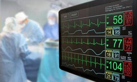
Monitoring during the initiation of NPPV in the elective setting should also be noninvasive. Monitoring is essential for heart rate, respiratory rate, use of accessory muscles of ventilation, ventilatory pattern, and particularly, perception of comfort and work of breathing. Generally, the successful application of NPPV is associated with a normalized ventilatory pattern and the patient’s perception of improved well-being. In addition, assessment of successful treatment requires long-term, close follow-up care. Regularly scheduled home visits are essential.24
In their initial NPPV treatment, patients fall into three categories: those who tolerate treatment well, understand the issues, and continue; those requiring additional sessions; and those who do not tolerate treatment at all. Prior to considering the patient intolerant of NPPV, however, every effort should be made to assist the patient. Often, a polysomnogram (PSG) may be required to titrate NPPV. Some PSG laboratories titrate NPPV and measure the work of breathing by placing a monitoring lead on the sternocleidomastoid muscles. NPPV settings are then titrated to decrease the work of this muscle, as evidenced by the readout.
Conclusion
ALS care remains treatment of the symptoms of the disease. A cure has not been found. Each case of ALS is as unique as the individual it affects, and care should be tailored to meet the needs of the individual and their family. The patient should have access to a multidisciplinary team of experts to evaluate his or her health status and therapies periodically. The multidisciplinary team must include a physician, a social worker, a nutritionist, a physical therapist, a nurse, and an RCP. Each discipline’s representative must have experience and a complete understanding of his or her role in the treatment of patients with ALS. More important, each clinician treating ALS should be cognizant of the array of options for each treatment decision and the implications that the decision may have for the outcome of that patient’s treatment. The RCP is a vital part of this team; however, like any other team member involved in the care of ALS, the RCP must have experience in treating individuals with neuromuscular diseases, as well as complete knowledge and understanding of therapeutic interventions and equipment capabilities. Requesting the initiation of noninvasive ventilation of an RCP lacking training and experience in using this treatment modality is like requesting bronchial lavage of an ophthalmologist. For home care, the implications can be far worse. Anyone with a DME license can sell respiratory equipment, including ventilators. RCPs experienced in the care of patients with neuromuscular disease should be in patients’ homes to treat them properly and monitor their progress for therapeutic success.
Acknowledgments
The author wishes to thank Gregory Ferriss, MD, and Penny Keller. She also wishes to give special thanks to all of her patients for their faith and trust.
Anna Chiappetta, BSRT, RRT, RCP, is New Orleans area supervisor for Health Management Services Inc, and president of AMC Consulting Services, both in New Orleans.
References
1. Walling AD. Amyotrophic lateral sclerosis: Lou Gehrig’s Disease. Am Fam Physician. 1999;59:1489-1496.
2. Cole P, Mitsumoto H. Amyotrophic lateral sclerosis. Clinical Neurology. 1998;4:1-27.
3. Annergers JF, Appel S, Lee JR, et al. Incidence and prevalence of amyotrophic lateral sclerosis in Harris County, Texas, 1985-1988. Arch Neurol. 1991;48:589-593.
4. Juergens SM, Kurland LT, Okazaki H, et al. ALS in Rochester, Minnesota, 1925-1977. Neurology. 1980;30:463-470.
5. Kutzke JF. Risk factors in amyotrophic lateral sclerosis. In: Rowland LP, ed. Amyotrophic Lateral Sclerosis and Other Motor Neuron Diseases. New York: Raven Press; 1991:245-270.
6. Brooks BR. Clinical epidemiology of ALS. Neurology Clinics of North America. 1996;14:399-420.
7. Chancellor AM, Hendry A, Caird FI, et al. Motor neuron disease: a disease of old age. Scott Med J. 1993;38:178-182.
8. Jackson CE, Bryan W. Amyotrophic lateral sclerosis. Semin Neurol. 1998;18:27-39.
9. Milonas I. Amyotrophic lateral sclerosis: an introduction. J Neurol. 1988;245:S1-S3.
10. Louvel E, Hugon J, Doble A. Therapeutic advances in amyotrophic lateral sclerosis. Trends Pharmacol Sci. 1997;18:196-203.
11. Carter GT, Miller RG. Comprehensive management of amyotrophic lateral sclerosis. Physical Medicine and Rehabilitation Clinics of North America. 1988;9:271-284.
12. Young JM, McNicoll P. Against all odds: positive life experiences of people with advanced amyotrophic lateral sclerosis. Health Soc Work. 1998;23:35-43.
13. Miller RG, Rosenburg JA. Practice parameter: the care of the patient with amyotrophic lateral sclerosis (an evidence-based review). Report of the Quality Standards Subcommittee of the American Academy of Neurology. Neurology. 1999:52:1311-1323.
14. Bach JR. Mechanical insufflation-exsufflation: comparison of peak expiratory flows with manually assisted and unassisted coughing techniques. Chest. 1993;104:1553-1562.
15. Chiappetta A, Beckerman R. High frequency chest wall oscillation in a patient with SMA: a case report. RT. 1995;8:112-114.
16. Aboussouan LS, Khan S. Effects of noninvasive positive pressure ventilation on survival in amyotrophic lateral sclerosis. Ann Intern Med. 1997;127:450-453.
17. Kacmarek B. Selection of a ventilator for the home. Presented at the 7th International Conference on Noninvasive Ventilation; March 15, 1999; Orlando, Fla.
18. Fallat RJ, Jewitt B. Spirometry in amyotrophic lateral sclerosis. Arch Neurol. 1979;36:74-80.
19. Cazzolli PA, Oppenheimer EA. Home mechanical ventilation for amyotrophic lateral sclerosis: nasal compared to tracheostomy-intermittent positive pressure ventilation. J Neurol Sci. 1996;139:S123-S128.
20. Smyth A, Riedi M. End of life decisions in amyotrophic lateral sclerosis: a cross-cultural perspective. J Neurol Sci. 1997;152:S93-S96.
21. Bach JR. Amyotrophic lateral sclerosis. Communication status and survival with ventilatory support. Am J Phys Med Rehabil. 1993;72:343-349.
22. Moss AH, Oppenheimer EA. Patients with amyotrophic lateral sclerosis receiving long-term mechanical ventilation: advanced care planning and outcomes. Chest. 1996;110:249-255.
23. Leger S. The choice of interface: can it make a difference? Presented at the 7th International Conference on Noninvasive Ventilation; March 16, 1999; Orlando, Fla.
24. Kacmarek R. Initiation of NPPV outside the ICU. Presented at the 7th International Conference on Noninvasive Ventilation; March 15, 1999; Orlando, Fla.
25. Hill N. Outcomes of NPPV in the hospital. Presented at the 7th International Conference on Noninvasive Ventilation; March 15, 1999; Orlando, Fla.