A new technique for cardiac magnetic resonance (CMR) imaging was shown to improve accuracy by removing patients’ need to breathe.
“In many imaging techniques, but particularly in CMR, you need a relatively long acquisition time and must correct for respiratory motion,” said Professor Juerg Schwitter. “For decades we have had to correct for respiration when estimating the position and motion of the heart by CMR, and this is not always accurate.”
He continued: “In Lausanne, radio-oncologists are exploring a technique called high frequency percussive ventilation. Patients do not need to breathe naturally and no correction for respiratory motion is required. This enables the physicians to more accurately plan the field of radiation to apply in each patient.”
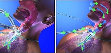
For the technique, patients put a mask over their mouth which is connected to a ventilator that delivers small volumes of air, called “percussions.” Instead of the 10 to 15 large breaths patients would take naturally per minute, air is provided in 300 to 500 small ventilations per minute. Air volumes are small so the chest does not move. The procedure is noninvasive, patients are conscious, not sedated, and do not need to breathe.
For the current study, the ventilator was adapted for use in the CMR environment. CMR uses a magnet which means that metal parts had to be swapped for MR-compatible ones. Tubes were used to connect equipment inside and outside the scanning room.
Professor Schwitter said: “Patients lie face up in the CMR machine and do not need to breathe. They say their chest feels a bit inflated during the ventilation but otherwise it feels okay.”


