These are images from endotracheal tubes: a) 5000× SEM image of 120-h postintubation endotracheal tube showing the presence of extracellular polymeric substrate; (b) 5000× SEM image of same endotracheal tube in a different location; (c) 15,000× SEM of same endotracheal tube focusing in on a cluster of cells; and (d) corresponding OCT image. doi:10.1117/1.JBO.20.12.126010
Researchers at the University of California Irvine demonstrated that optical coherence tomography (OCT) can be used to determine the presence of biofilm — bacterial growth that results from the attachment and subsequent multiplication of microorganisms to a synthetic surface.
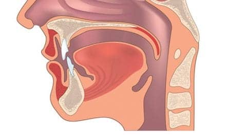
Biofilm has been linked to ventilator-associated pneumonia (VAP), which is a prevalent infection in hospital intensive care units. Using OCT, the team was able to visualize and assess the extent of airway obstruction from biofilm deposited on intubated endotracheal tubes in vivo.
According to the study published in the Journal of Biomedical Optics, extubated patients’ endotracheal tubes were imaged using both OCT and scanning electron microscopy (SEM). Biofilm was present in both images, and the researchers were able to use the OCT image to measure a reduction in endotracheal inner lumen area between a 1-day intubation sample tube and a 5-day sample tube.
The study showed that OCT can be used to both detect the presence of biofilm and gather further information about the extent of biofilm formation from the thickness of biofilm observed.