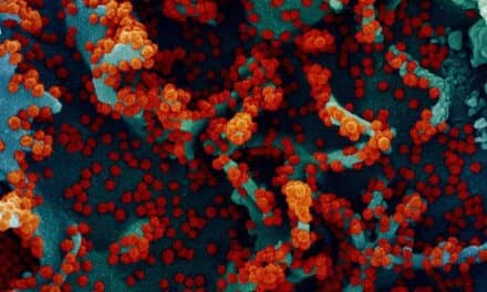
A new study of the monkeypox virus has revealed how the virus might damage lungs during infection. Not only does the infection from the monkeypox virus increase production of proteins involved in inflammation, but it decreases production of proteins that keep lung tissue intact and lubricated. The findings, published in the journal Molecular & Cellular Proteomics, also suggest possible new ways to treat lung diseases in humans.
“Going into this study, we thought monkeypox caused disease primarily by inducing inflammation in the lung, and that leads to pneumonia,” said Joseph Brown, lead author and a systems biologist at the Department of Energy’s Pacific Northwest National Laboratory (PNNL). “We were surprised to see how badly the virus wrecked the structural integrity of the lungs.”
Monkeypox and smallpox are closely related viruses that cause contagious pustules in humans, although monkeypox is less dangerous. Monkeypox is as bad for monkeys as smallpox is for people, however, making it a good model for human smallpox disease. The study helps researchers better understand both monkeypox and smallpox infection.
“If researchers confirm similar events in people, doctors might be able to give surfactants—lubricating chemicals that aid in gas exchange—to help the lung function. And the findings could lead to new areas of pulmonary studies in general—bronchitis or emphysema, lung transplants, the flu,” said Josh Adkins, also from PNNL.
Monkeypox infections in humans have been on the rise since smallpox was eradicated in the late 1970s. Up to 10% of those infected with monkeypox die of the disease. Monkeypox can be caught from infected rodents, pets, and monkeys. Although mainly found in Africa, the first documented infection in the United States occurred in 2003.
Few studies exist that look at how monkeypox infection damages the lungs. Because symptoms in macaque monkeys and humans are so similar, the research team examined how the virus affected the collection of proteins found in lung fluid from macaques. The researchers infected the macaques with the monkeypox virus and followed the course of infection in the lungs of individual animals. They then washed the lungs of the infected monkeys with a saline solution and analyzed the washes for protein analysis. The complement of proteins produced by lung tissue before and during infection would indicate how the lungs are responding to the virus.
Early in infection, monkeypox virus simulated an immune response, ratcheting up production of proteins associated with inflammation. The monkeypox-infected lungs, however, also showed a distinct decrease in some proteins—proteins involved in metabolism, structural proteins that serve as I-beams and cross-beams of lung tissue, and finally surfactant proteins, which provide lubrication and help with oxygen exchange. Inflammatory proteins decreased over time in the monkeypox infected lung fluid samples, but the structural proteins continued to stay low.
“Our results suggest that inflammation contributes to disease but it may not be the main component. Interfering with the structural proteins may play a major role,” said Brown.
The researchers found similar trends with cultured cells, suggesting that some aspects of the monkeypox infection can be studied in test tubes. In addition, the team directly detected viral proteins in the lung fluid samples. Usually, scientists needs to use antibodies to detect viral proteins because there are so few of them swimming in a sea of host proteins.
Ultimately, this type of research could have wider implications than viral infection. “This study serves as a great reference for pulmonary diseases,” said Adkins. “It opens up the doors for other lung fluid studies.”
Source: Pacific Northwest National Laboratory


