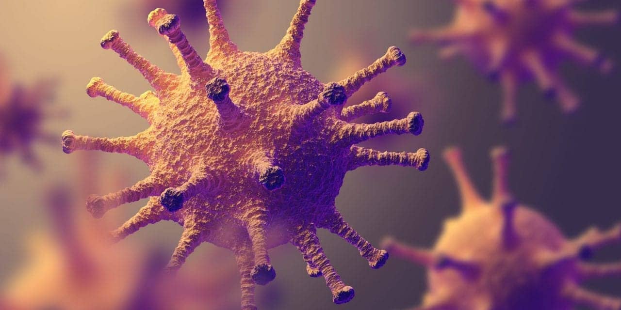
The influenza A virus (IAV) remains in heart tissue after it has been cleared from the lungs, according to a murine study published in Circulation Research by scientists from the J. Craig Venter Institute.
Live IAV present during convalescence does not actively replicate, preventing development of antiviral inflammatory responses, thus cloaking it from the immune system. Undetected, the virus continues to disrupt mitochondrial function, causing a metabolic breakdown and promoting cell death, the researchers say.
“Recent studies have pointed towards a direct link between influenza patients and later cardiac events, so we set out to try and understand the molecular mechanisms involved. While we expected to find myocardium (heart muscle) damage, we did not expect to find active IAV during convalescence,” said senior author and JCVI assistant professor Norberto Gonzalez-Juarbe, PhD.
Wild-type and MLKL deficient mice—which lack the ability to undergo programmed necrosis, ie necroptosis—were infected with either the “2009 pandemic” strain or a laboratory Influenza A strain. The present study focused on the pandemic strain as previous clinical reports showed a higher rate of adverse cardiac events during each of the last influenza pandemics.
Once the infection had cleared the lungs, specimen heart tissue was analyzed using immunofluorescence microscopy and plaque assays which showed the presence of viable IAV particles in the myocardium. Further analysis on the global proteome and phosphoproteome was performed using mass spectrometry. This showed that both the proteome and phosphoproteome were significantly altered in IAV-infected mouse hearts, regardless of strain.
The proteome is the complete complement of proteins that can be expressed whereas the phosphoproteome is a subset of proteins that contain a phosphate group used in cell signaling networks. Disrupting cell signaling interferes with cellular function, metabolism, and immune responses.
The necroptosis deficient mice had increased survival and reduced weight loss post-IAV infection, as well as increased antioxidant and mitochondrial function, indicating partial protection to IAV infection. These observations suggest inhibition of necroptosis or prevention of mitochondrial damage as possible therapeutic interventions to reduce cardiac damage during influenza infections.
Given these findings, additional research is warranted on the effects of other pulmonary pandemic viruses on cardiac health. This is even more relevant given the current widespread observations of adverse cardiac events in COVID-19 patients.