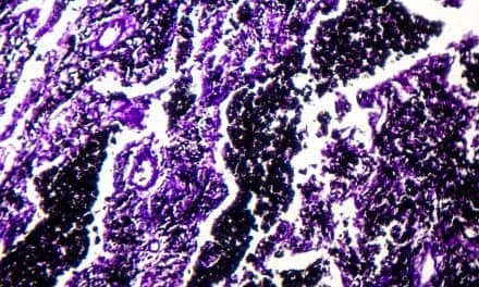
A new study finds a large number of individual sites within the lungs undergo active disease scarring in pulmonary fibrosis. Previously, scientists believed the scarring progressed like a wave from the outside to the inside of the lung.
To make this discovery, Southampton Respiratory Biomedical researchers used advanced 3D X-ray imaging technology originally designed for the analysis of engineering parts, such as jet turbine blades. Insights gained from this powerful scanning equipment should help scientists develop targeted therapies focusing on these areas, the researchers said.
Within each tissue sample, researchers saw numerous, complex fibroblasts that varied in their shape and volume. Fibroblasts are cells that synthesize collagen and proliferate in pulmonary fibrosis, contributing to the thickening of lung tissue. Unexpectedly, however, each fibroblast was an independent structure with no connections to other fibroblasts.
New 3D-Imaging Technique Reveals How Pulmonary Fibrosis Develops