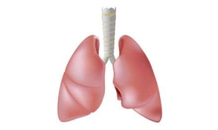
A new study published in Nature Communications shows that bacteria are often present deep inside the lungs of COPD patients. With a 2-year grant from the American Lung Association, Vanderbilt University researchers investigated the function of the body’s mucus membranes, such as those found in the lining of the lungs. According to an HCP Live report, one belief about COPD development is that inhaling toxic gases and particles from tobacco smoke causes the inflammation in small airways; however, the authors of the study say this theory does not explain why the disease continues to progress even after smoking cessation.
The research team notes that these surfaces typically have barriers that prevent bacteria from penetrating deep into the tissue of those organs, but the study’s authors previously showed that COPD patients frequently lack this barrier, called secretory immunoglobulin A (secretory IgA), and used mice lacking secretory IgA to test their hypothesis.
The HCP Live report indicates that the mice appeared healthy when they were born, but as they got older, the mice developed increased bacterial invasion, chronic inflammation, and damage to their lungs mimicking human patients with COPD, the researchers reported. These changes often were present without exposure to cigarette smoke or other toxin exposure.
The research team was able to stop the damage in the secretory IgA mice by using the anti-inflammatory drug roflumilast. The researchers believe that this suggests that COPD patients with decreased secretory IgA in their small airways could one day use roflumilast to treat their increased bacterial invasions, as indicated on the HCP Live report.
First author of the study Bradley Richmond, MD, says, “These findings suggest that over time changes in the airways resulting from cigarette smoke exposure make the lungs more susceptible to bacterial invasion. This may explain why inflammation persists in COPD even after patients stop smoking.”
Source: HCP Live