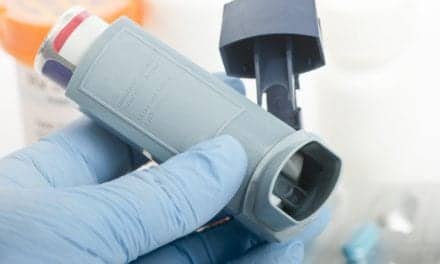
Helium-oxygen mixtures have proven very effective in treating hospital respiratory emergencies
At midday, February 7, 1998, a 26-year-old woman arrived at the emergency department (ED) of Wesley Medical Center, in Wichita, Kan, via emergency medical services (EMS) transport. She was in respiratory failure secondary to suspected status asthmaticus. The patient had been cooking lunch when she became severely short of breath. She had a 2- to 3-day history of some shortness of breath, but in general had been feeling fairly well. She had been using her inhalers and had noticed some itching around her neck and throat. The patient said this was a common occurrence, and had taken diphenhydramine hydrochloride, as well as aspirin, for a headache the day before.
On this day, she had not taken any medications by mouth. Her symptoms went from baseline to maximal in approximately 1 hour, according to her husband. EMS personnel were summoned, and they coded the patient red. Epinephrine was requested and administered at the scene. Shortly afterward, EMS personnel reported a run of bradycardia and subsequent pulseless electrical activity lasting 5 to 10 seconds. The patient’s pulse then resumed spontaneously. En route to the hospital, intubation was attempted, but EMS personnel were unable to place the endotracheal tube because of suspected laryngeal swelling. They reported that the patient was very cyanotic and that breath sounds were completely absent.
Assessment and History
The patient had a history of asthma, starting at age 4, but said she did not have any other major illnesses. She was using fexofenadine, albuterol sulfate, and triamcinolone acetonide, and she had a significant history of using steroids, although she had not used them recently. The patient also stated she did not use any recreational drugs. She had multiple allergies to many environmental allergens, but no known drug allergies. Several days before the episode, she had completed a 3-day course of trimethoprim sulfate and sulfamethoxazole for a urinary tract infection.
On arrival at the ED, the patient had an oxygen saturation, as measured using pulse oximetry (Spo2), of 81 percent while being ventilated via bag-mask combination with 100 percent oxygen. She was negative for fever, nausea, and vomiting. Her pulse rate was 110 to 118 beats per minute. She was in marked distress and entirely cyanotic, with absent breath sounds. She was slightly combative and not verbally responsive. Anesthesia staff were standing by because of the difficulty that EMS personnel had encountered in attempting to intubate the patient.
There was neither mucosal swelling nor obstruction in her upper airway. Initial blood gas results, from a sample obtained during bag-mask ventilation with 100 percent oxygen, showed a pH of 7.07, a Pco2 of 81, a Po2 of 250, a bicarbonate (HCO3) level of 17.5, and a base excess (BE) of -8.8. Her white blood cell count was 15.9. Her skin was cool and blue, with good capillary perfusion. She was negative for any airway edema. Peripheral pulses were present. Her chest radiography findings were negative. The initial overall patient impressions of the ED staff were of status asthmaticus, status postcardiac arrhythmia, recent urinary tract infection, and multiple environmental allergies.
Treatment
In addition to the bag-mask combination using 100 percent oxygen, the patient, on arrival at the ED, also received continuous albuterol breathing treatments with a mixture of 80 percent helium and 20 percent oxygen powering the nebulizer (which was connected by a T-tube to the bag-mask combination). She was also given methylprednisolone intravenously. Relief was quickly noted as the patient went from absent breath sounds through wheezing to fairly good airflow within minutes. As the patient’s condition quickly improved and her Spo2 levels increased, the bag’s gas supply was changed to a helium-oxygen mixture and the nebulizer’s supply was changed to 100 percent oxygen. The patient was rapidly improving and was stable. Once she was making spontaneous respiratory efforts, she was given an 80 percent helium and 20 percent oxygen mixture via nonrebreathing mask with a 3 L/min oxygen flow delivered via nasal cannula.
Continuous albuterol treatments were administered with the helium-oxygenmixture. The patient’s Spo2 improved to 92 percent to 94 percent, and intubation was no longer indicated. Bag-mask ventilation was continued for approximately 10 to 15 minutes until the nonrebreathing mask was applied. The patient was transferred to the medical intensive care unit while using the same gas flows and receiving continuous albuterol. She was asleep at the time of transfer, with a respiratory rate of 12 breaths per minute.
Course of Hospitalization
Approximately 2.5 hours after admission to the ED, an arterial sample taken while the patient was being given supplemental oxygen at a rate of 3 L/min yielded these results: pH, 7.43; Pco2, 30; Po2, 90; HCO3, 21.5; and BE, -3.6. The patient continued to improve and was weaned from the helium-oxygen mixture and the continuous albuterol treatment 4 hours after her arrival. Two and a half hours later, another blood gas analysis was done; pH and Pco2 values were similar, but there was an improved Po2 (of 140) with no changes in oxygen flow rates. The patient’s albuterol breathing treatments were decreased to every 4 hours, and intravenous methylprednisolone was continued.
The patient had probably had an anaphylactic reaction (versus status asthmaticus). It was speculated that this could have been caused by a combination of sulfa drugs and aspirin. The patient was discharged without wheezing approximately 48 hours after admission to the ED.
Helium-oxygen mixtures
This case is yet another strong demonstration of a helium-oxygen mixture’s value in the treatment of upper-airway obstruction. These mixtures were first used in 1934 by Barach1 to treat obstructive lesions of the airway. The use of, and interest in, helium continued into the early 1940s. Helium was largely abandoned until the late 1980s, when it was used to treat 10 patients with status asthmaticus and subsequent respiratory acidosis. These patients were treated with aerosolized bronchodilators and intravenous aminophylline. Several of the patients required subcutaneous epinephrine. The results of initial blood gas analyses for all patients revealed either respiratory acidosis or combined metabolic and respiratory acidosis. After helium-oxygen mixtures containing 25 percent to 40 percent oxygen were administered, prompt resolutions of symptoms were noted.1 None of the patients required intubation, and it was concluded that improved alveolar ventilation promotes rapid washout of alveolar carbon dioxide and enhanced removal of arteriolar carbon dioxide.1 Scattered reports of the use of helium-oxygen mixtures have been seen in the medical literature since that time.
The physical properties of helium make it useful for the treatment of respiratory conditions such as upper-airway obstruction, status asthmaticus, severe chronic obstructive pulmonary disease, croup, postextubation stridor, and high-frequency jet ventilation. It has also had very profound effects on infants with respiratory syncytial virus infection secondary to bronchiolitis. Helium is seven times lighter than nitrogen, so replacing the nitrogen found in room air with helium produces a gas with a density one-third that of air. Its benefits are threefold. First, with its low density, it results in a much less turbulent flow in narrowed airways. A decrease in airway resistance will result in decreases in work of breathing and oxygen consumption. Second, helium has the ability to eliminate carbon dioxide four times more quickly than a nitrogen-oxygen mixture. Third, gas distribution throughout the lungs is improved because of better alveolar ventilation.
There are some potential drawbacks to helium therapy, however. Helium has a high thermal conductivity, and there is a risk of hypothermia when the temperature of the helium mixture is less than 36øC. Mixtures are available in concentrations of 60 percent to 80 percent helium; these are very suitable for ED use. All commercial mixtures contain at least 20 percent oxygen to prevent hypoxia. In this case, supplemental oxygen was used to increase the fraction of inspired oxygen. Caution must be exercised, however; too high an oxygen concentration will cause the gas mixture to become dense and lose its efficacy. If the patient presents with acute hypoxia, a helium-oxygen mixture may be contraindicated if adequate oxygenation cannot be maintained.
Helium-oxygen mixtures are available in portable cylinders, making transport very simple. Because helium is such a highly diffusible gas, a tight fit should be maintained between the patient and the mask. Lower flows can be used to keep the reservoir bag inflated because of the low specific gravity of helium. Helium is an inert gas that has not been known to interact with human metabolism, so it is safe for all patients. Pulse oximetry should always be performed during the administration of helium-oxygen mixtures because patients with high oxygen requirements may not tolerate high concentrations of helium. Helium-oxygen mixtures produce no cure on their own; therefore, treatment of the underlying abnormality must be accomplished.
Conclusion
Helium-oxygen mixture use has proven to be a very effective therapy for many different types of hospital respiratory emergencies. RCPs should familiarize themselves with the use of these mixtures and should consider their use until adjunctive care for the patient can be provided. By decreasing or eliminating the need for mechanical ventilation, better patient outcomes and shorter hospital lengths of stay can be obtained.
Steven B. Snook, RRT, is a perinatal-pediatric specialist at Wesley Medical Center, Wichita, Kan.
Acknowledgement
The author would like to thank Gloria Jones, RRT, for her assistance.
REFERENCE
1. Shiue ST, Gluck EH. The use of helium-oxygen mixtures in the support of patients with status asthmaticus and respiratory acidosis. J Asthma. 1989;26:177-180.