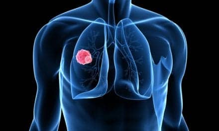
Recent advances in gene therapy offer new hope for successful treatment of lung cancer.
 Each year, there are an estimated 180,000 new cases of lung cancer and 160,000 deaths from lung cancer in the United States.1 Despite the introduction of new chemotherapeutic agents, refinements in surgery, and advances in radiation-therapy techniques, survival rates for lung cancer have remained relatively unchanged; the overall survival rate is still less than 13%.1 There has been some improvement in results when multimodality treatments (such as primary chemotherapy plus radiation therapy, or surgery to reduce the tumor burden followed by chemotherapy and/or radiation therapy) have been employed, but the toxicities of multimodality treatments sometimes limit their usefulness.
The identification of specific genetic abnormalities that contribute to the process of carcinogenesis has provided an opportunity to develop cancer treatments aimed at reversing those genetic abnormalities. The concept of gene therapy is based on the observation that certain diseases are caused by the inheritance of a single functionally defective gene. Replacement of the defective gene theoretically could treat—and possibly cure—the disease. Unfortunately, gene therapy for lung cancer is more complicated because multiple genetic defects are usually present, and the replacement of all defective genes is not possible using current gene-transfer techniques.
Lung Cancer Development
Multiple genetic mutations are required to transform a normal lung cell into a cancer cell. These mutations cause activation of oncogenes (dominant cellular factors that stimulate a cell to divide) and deletion of tumor-suppressor genes. These genetic changes result in pathological states that are reversible in their early stages (see Figure 1). As progressive chromosomal changes become more complex, however, lung cancer develops.
|
| Figure 1. Sequence of genetic events in lung cancer: |
Lung cancer results not only from abnormal cell proliferation, but also from abnormalities in the intrinsic cell-death program of the cell (apoptosis). Tumor mass is a balance between cell proliferation and cell death. Resistance to chemotherapy may be due, in part, to deficits in the cell-death program, resulting in an increasing population of chemoresistant lung-cancer cells. Such processes are regulated by factors both intrinsic and extrinsic to the cell, including growth factors, cytokines, and hormones. These factors interact with specific receptors and communicate with the nucleus through intracellular signaling pathways, resulting in the activation of genes associated with proliferation and the inhibition of those associated with apoptosis.
Oncogenes in lung-cancer cells code for mutated proteins or for the overexpression of proteins involved in the pathways that signal proliferation and apoptosis. The pathways are thereby subverted, allowing the tumor to grow. Lung-cancer cells may also manipulate their local environment to facilitate tumor growth, particularly by suppressing the host’s immune response and promoting the development of new blood vessels (angiogenesis).
Gene-therapy strategies proposed for the treatment of lung cancer include modulation of the immune system, suicide-gene therapy, inhibition of oncogenes or replacement of tumor-suppressor genes, and enhancement of conventional chemotherapy and radiation therapy.1
Modulation of the Immune System
According to Jack A. Roth, MD, chief of the Thoracic Molecular Oncology Section, Department of Thoracic and Cardiovascular Surgery, University of Texas M.D. Anderson Cancer Center, Houston, “Various strategies have been proposed for enhancing the immune system. In animal models, tumor regression can be demonstrated after administration of various cytokines, including interleukin (IL)-2, IL-4, IL-6, IL-7, IL-12, granulocyte-macrophage colony-stimulating factor, granulocyte colony-stimulating factor, tumor necrosis factor (TNF)-a, and a-interferon (IFN), and g-IFN.”
Roth notes, however, that the systemic administration of cytokines in human trials has been limited by their toxicity and by incomplete response at the tumor site. Gene-therapy strategies, therefore, have been proposed to minimize these systemic side effects and increase tumor response. Cytokine-based strategies include the development of tumor-cell vaccines that incorporate genetically modified fibroblasts or tumor cells that secrete cytokines. Mice vaccinated with radiation-inactivated Lewis lung carcinoma cells transfected with various cytokines, including macrophage colony-stimulating factor, IL-2, and TNF-a, develop improved antitumor immunity to subsequent tumor inoculation. Roth says, “Investigators have demonstrated that cytokines act in a paracrine (rather than systemic) fashion, and have tried to enhance local cytokine activity by injecting genetically engineered fibroblasts or tumor cells intratumorally. For example, direct injection of IL-12–engineered fibroblasts into flank tumors in mice resulted in long-term protective antitumor immunity to pulmonary metastases. Other studies have shown that the intrapleural administration of IL-2—transfected lung-cancer cells in mice has resulted in local tumor regression and long-term antitumor immunity.”
Roth adds, “Clinical trials that involve the adoptive transfer of IL-2–activated lymphocytes (lymphokine-activated killer cells or tumor-infiltrating lymphocytes) and systemic IL-2 therapy have shown therapeutic promise in patients with renal-cell carcinoma and melanoma, but only minimal activity in lung cancer. Efforts to increase the antitumor activity of these lymphocytes have included the genetic transfer of cytokines into the lymphocytes.”
Another approach to making tumor cells more immunogenic is to transfer costimulatory molecules such as B7-1 and B7-2, which are often lacking on the tumor-cell surface. Such molecules are needed for the effective recognition of tumor antigens by the immune system. One potential difficulty with this approach, however, is the heterogeneity and unpredictability of costimulatory gene expression in tumors. Therefore, Roth says, “This approach may require replacement of multiple genes to elicit an effective immune response.”
Suicide-Gene Therapy
Suicide-gene therapy involves the transduction of tumor cells with a gene capable of converting a nontoxic compound (prodrug) into a toxic metabolite. The transferred suicide gene then selectively kills the transduced cell after systemic administration of the prodrug. The two most commonly used genes for suicide-gene therapy are the herpes simplex virus–thymidine kinase (HSV-tk) gene and the cytosine deaminase gene. HSV-tk converts nontoxic ganciclovir to a cytotoxic triphosphate metabolite. Cytosine deaminase converts 5-fluorocytosine to the cytotoxic antimetabolite 5-fluorouracil. Tumor-killing activity is enhanced in both systems by a bystander effect in which toxic metabolites are transferred from transduced to nontransduced cells through intercellular gap junctions.
Inhibition or Replacement of Critical Genes
Roth explains that the identification of specific genes critical to the development of lung cancer offers an opportunity to target these genes or their products for treatment. The genes implicated in carcinogenesis include dominant oncogenes and mutated or deleted tumor-suppressor genes. Oncogenes develop from proto-oncogenes (normal cellular homologues of genes normally involved in critical cell functions such as signal transduction and transcription). Point mutations, amplifications, translocations, or rearrangements can change a single allele of a proto-oncogene into a dominant transforming oncogene. “Tumor-suppressor genes, on the other hand, require the inactivation of both alleles of the gene before transformation of the cell,” Roth says. “Certain oncogenes and tumor-suppressor genes may be critical to the development of the malignant phenotype. Thus, gene-therapy strategies that inactivate critical oncogenes or replace critical tumor-suppressor genes may lead to tumor regression by reversion of the malignant phenotype.”
Oncogenes of the ras family are some of the most commonly activated oncogenes in lung cancer, and are therefore potential targets for oncogene-inactivating strategies. Roth says, “In preclinical studies, transduction of lung cancer cells with an antisense K-ras complementary DNA plasmid has been shown to block selectively the production of mutant K-ras messenger RNA and to reduce the growth of lung cancers in vitro and in vivo in mice.” Antisense DNA is the strand of DNA complementary to the one bearing the genetic message, and the one from which it may be reconstructed. Roth adds, “Similarly, treatment of murine lung-cancer cells with an antisense cyclin D1 construct reduced in vitro and in vivo proliferation and tumorigenicity, presumably by inhibiting expression of the cyclin D1 oncogene.”
Another strategy aimed at inhibition of an oncogene involves the transfer of a gene whose expression is known to block the activated oncogene. The HER-2/neu oncogene encodes an epidermal growth factor-related transmembrane protein (p185) with intrinsic tyrosine kinase activity. “Amplification or overexpression of HER-2/neu has been demonstrated in many solid tumors, and has been correlated with poor prognosis and chemoresistance in lung cancer,” Roth says. “The adenovirus type 5 early region 1A gene product can suppress HER-2/neu–mediated malignant transformation by inhibiting HER-2/neu expression.”
Tumor-suppressor genes are a class of genes that are important in growth regulation and whose absence may contribute to tumor growth. The p53 gene is one of the most commonly mutated genes in lung cancer, and is believed to be a critical tumor-suppressor gene.2 One of the most intriguing approaches to the gene therapy of lung cancer involves the introduction of the p53 tumor-suppressor gene into tumor cells. The thoracic surgery group at the University of Texas M.D. Anderson Cancer Center, headed by Roth, has conducted a series of experiments using wild-type p53 constructs delivered by retroviral or adenoviral vectors to affect tumor-cell growth.3-7 Transduction of wild-type p53 into lung cancer cell lines with deleted or mutated p53 increased sensitivity to the antitumor drug cisplatin.6
Enhancement of Conventional Therapy
According to Roth, “Chemotherapy and radiation therapy induce tumor-cell death, in large part, by causing DNA damage that leads to apoptosis of the tumor cell. The molecular mechanisms underlying this process may involve wild-type p53. Cellular p53 protein levels rise in response to DNA damage and induce gap 1 (G1) arrest, which allows the cell to repair its DNA before entry into S phase,” the synthesis period that follows G1 (the gap in the somatic cell cycle that follows mitosis) and precedes G2. “If the DNA is not repaired,” Roth continues, “apoptosis may be induced through a p53-dependent pathway. Cells with mutated or deleted wild-type p53 genes appear to be resistant to chemotherapy-induced or radiation-therapy–induced apoptosis, whereas cells in which wild-type p53 protein is restored by gene transfer appear to be more sensitive.”
The administration of adenoviral vectors with wild-type p53 in lung-cancer cells before chemotherapy has resulted in enhanced apoptosis, both in vivo and in vitro. Similar results have been noted in colon-cancer cells after radiation therapy, Roth reports. Because of these encouraging results, a phase I clinical trial in patients with advanced non–small-cell lung cancer has begun at the University of Texas M.D. Anderson Cancer Center. If these genes are ultimately shown to increase chemotherapy-induced or radiation-therapy–induced cell death without increasing toxicity to normal tissues, they may prove to be effective adjuncts to conventional treatments.
Conclusion
Gene therapy for lung cancer is still in its infancy. Although few patients have been treated, early results are promising. As the technology of gene therapy advances, new opportunities will be available to initiate lung-cancer gene-therapy trials that are based on a more detailed understanding of the molecular biology of lung cancer. It is hoped that the future will bring more targeted, more effective, and less toxic methods to prevent the development and progression of lung cancer.
John D. Zoidis, MD, is a contributing writer for RT Magazine.
References
1. Swisher SG, Roth JA. Gene therapy for human lung cancers. Surg Oncol Clin N Am. 1998;7:603-616.
2. Carbone DP, Minna JD. The molecular genetics of lung cancer. Adv Intern Med. 1992;37:153-171.
3. Cai DW, Mukhopadhyay T, Liu Y, et al. Stable expression of the wild-type p53 gene in human lung cancer cells after retrovirus-mediated gene transfer. Hum Gene Ther. 1993;4:617-624.
4. Zhang WW, Fang X, Mazur W, et al. High-efficiency gene transfer and high-level expression of wild-type p53 in human lung cancer cells mediated by recombinant adenovirus. Cancer Gene Ther. 1994;1:5-13.
5. Fujiwara T, Grimm EA, Mukhopadhyay T, et al. A retroviral wild-type p53 expression vector penetrates human lung cancer spheroids and inhibits growth by inducing apoptosis. Cancer Res. 1993;53:4129-4133.
6. Fujiwara T, Grimm EA, Mukhopadhyay T, et al. Induction of chemosensitivity in human lung cancer cells in vivo by adenovirus-mediated transfer of the wild-type p53 gene. Cancer Res. 1994;54:2287-2291.
7. Liu TJ, Zhang WW, Taypor DL, et al. Growth suppression of human head and neck cancer cells by the introduction of a wild-type p53 gene via a recombinant adenovirus. Cancer Res. 1994;54:3662-3667.