RCPs’ knowledge of arterial blood gas results is key in helping patients regain their baseline health and avoid expensive therapies and unnecessary procedures.
By Bill Pruitt, CPFT, RRT, MBA
Arterial blood gases (ABGs) are considered to be the most valuable measurement available to assess the function of the cardiopulmonary system. They provide a wealth of information concerning oxygenation, ventilation, and acid-base status in order to guide the care and treatment of patients and are indispensable for the management of the critically ill from neonates to geriatrics. They provide a baseline measurement as well as track movement toward restored cardiopulmonary health or toward cardiopulmonary failure. Other measurements such as transcutaneous gas monitoring, pulse oximetry, and end-tidal carbon dioxide monitoring provide pieces of information that approximate some of the details used in assessing the cardiopulmonary system, but the blood gas is still the gold standard, giving the full complement of measured values with the highest degree of accuracy. This article will review the basics in obtaining arterial samples and provide a practical approach to interpreting ABG results.
Obtaining Arterial Samples
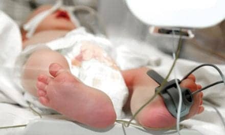
When arterial samples are obtained using a percutaneous needle puncture (in adults), the radial, brachial, or femoral arteries are the most commonly used sites, with the radial being the most preferred site. For infants, the most common sites to obtain an ABG are the radial and temporal (scalp) arteries. Other sites include the axillary, ulnar, and dorsalis pedis arteries, plus the umbilical artery (during the first 24 to 48 hours after birth). An indwelling arterial catheter allows for obtaining multiple samples without having to perform repeated percutaneous punctures (and allows for continuous arterial pressure monitoring).
Supplies needed for the arterial sample obtained by a percutaneous puncture or “arterial stick” include a heparinized syringe with a needle, alcohol wipes, gauze, bandage or tape, gloves, label, syringe cap, and needle cork to cover the needle tip. Supplies needed for drawing an arterial sample from an indwelling line include a heparinized syringe, gloves, label, and syringe cap. If the arterial sample is not going to be analyzed within 15 minutes of being drawn, an ice slurry is needed to store the sample by immersing the syringe barrel. This will slow the ongoing metabolism of the blood in the syringe and help assure that the results are a reliable reflection of the patient’s status at the time the sample was obtained. Most hospitals are using preheparinized syringes. For adults, a 20 to 22 gauge needle is used; for infants, a 25 gauge needle is used, and the needle length is from 5/8″ to 1.5″ long.
The procedure for obtaining a sample by arterial stick starts with checking a patient’s medical record for an order, then gathering the needed supplies. After proper handwashing, gloves are put on, and the patient’s identification is checked. For a radial artery stick, a modified Allen’s test is performed to assure that there is sufficient circulation through the ulnar artery in case something happens to interfere with radial artery perfusion. The area of the stick is thoroughly cleaned with the alcohol wipe, then the artery is palpated by feeling for the blood pulsation through it. The needle is inserted at a 45° angle and with an auto-fill syringe; the blood pressure will fill the syringe barrel and stop when the blood reaches the plunger stopper. The needle is withdrawn quickly and pressure is held over the site for 5 minutes (longer holding time is needed if the patient is on anticoagulant therapy) until the site is not oozing and a bandage or taped gauze covering is applied. The syringe is checked for air bubbles, which are removed if present, the needle is properly corked, and the sample is gently rolled or inverted to mix the blood with the anticoagulant. The sample is labeled with the date, time of sample, Fio2 or supplemental oxygen flow, and mode of oxygen delivery or mode of supported ventilation. Then the sample is taken to be analyzed immediately (within 15 minutes) or put in the ice slurry for later analysis (no longer than 60 minutes).
Interpreting the Results
After the sample has been analyzed on the blood gas machine, the results must be interpreted. The foundation for interpretation rests on the normal values and what is sometimes called the absolute values. Consider blood pressure: normal values for blood pressure would be: systolic 100-140 mm of mercury (mm Hg) and diastolic 60-90 mm Hg with an absolute normal of 120/80 mm Hg. In a similar way, blood gases have normal values and absolute normal values. Keep in mind that the measured results are influenced by many factors from pulmonary health to kidney function, and from disease states to the care that respiratory therapists give—particularly oxygen therapy and artificial ventilation. The ABG results should be considered in light of the influencing factors, while remembering that the results in hand are only a snapshot of the patient’s condition at the time that the sample was obtained. Consider that even the process of obtaining a sample from an arterial stick can cause patients to hyperventilate and change the values.
Acid-Base Status and Ventilation
The hardest part of interpreting an ABG results is figuring out the acid-base status; however, if certain questions are asked and answered in a step-wise fashion, the interpretation becomes easier. While moving through this process, keep in mind the following guidelines:
- An increased pH (for example, 7.43) indicates a move toward an alkalotic state (alkalosis).
- A decreased pH (7.36) indicates a move toward an acidic state (acidosis). Once the pH has crossed the limits of the normal range, the interpretation will clearly be either an alkalosis or an acidosis.
- An increased Hco3- (26 mEq/L) indicates a possible* move toward an alkalotic state (alkalosis) with a metabolic cause. A quick and easy way to remember this is to think of Hco3- as a base, which is a substance that takes H+ ions out of solution and increases pH.
- A decreased Hco3- (for example, 22 mEq/L) indicates a possible* move toward an acidic state (acidosis) with a metabolic cause. Recall Hco3- is a base substance, and loss of a base causes an increase in H+ ions (which is the same as a decrease in pH).
- An increased Paco2 (45 mm Hg) indicates a possible* move toward an acidic state (acidosis) with a respiratory cause. Think of Paco2 as an acid (a substance that increases H+ and decreases pH)
- A decreased Paco2 (35 mm Hg) indicates a possible* move toward an alkalotic state (alkalosis) with a respiratory cause.
The interpretation for an ABG will be defined by four elements: the acid-base status (either normal, an alkalosis, or an acidosis); the primary cause of the abnormality (either a respiratory or a metabolic cause or a combined cause in both respiratory and metabolic functions); whether or not the body is compensating for the abnormality (either not compensated, partially compensated, or fully compensated); and an assessment of oxygenation (from normal to mild, moderate, or severe hypoxemia).
Moving Through the Process
First, compare the pH to the absolute normal of 7.40. If the measured value is higher, then the patient may have an alkalosis (there is also the possibility that the acid-base status is normal if the pH is between 7.40 and 7.45). If the pH is above 7.45 (the high end of the range), the interpretation is definitely an alkalosis. If the pH is below the absolute normal of 7.40, the patient may have an acidosis (there is also the possibility that the acid-base status is normal if the pH is between 7.40 and 7.35). If the pH is below 7.35 (the low end of the range), the interpretation is definitely an acidosis.
Next, examine the results for the Paco2 and the Hco3- to determine the cause. If one of these is moving toward the same condition as the pH, this is labeled the primary cause. For example, given a pH of 7.46, a Paco2 of 33 mm Hg, and a Hco3- of 23 mEq/L, what is the interpretation of the pH and the primary cause? The pH is labeled as an alkalosis, the Paco2 is also alkalotic, and the Hco3- is normal (moving slightly off the absolute of 24 mEq/L). This would be called a respiratory alkalosis and reflects an increase in ventilation that is “blowing off” CO2 (better known as hyperventilation).
Given a pH of 7.46, a Paco2 of 39 mm Hg, and a Hco3- of 27 mEq/L, what is the interpretation of the pH and the primary cause? Again, the pH is labeled as an alkalosis, but the Paco2 is in the normal range and the Hco3- is alkalotic. This would be called a metabolic alkalosis. Some causes of this would be vomiting (or a nasogastric tube connected to a continuous suction), hypokalemia (sometimes caused by potassium-wasting diuretics such as furosemide), and excessive sodium bicarbonate administration. What if the pH was 7.50, the Paco2 was 24 mm Hg, and the Hco3- was 28 mEq/L? The pH is alkalotic and both the Paco2 and the Hco3- are alkalotic. This would be a combined metabolic and respiratory alkalosis.
Conversely, given a pH of 7.22, a Paco2 of 55 mm Hg, and a Hco3- of 25 mEq/L, what is the interpretation? Both the pH and the Paco2 reflect an acidic situation. The Hco3- is slightly off the normal of 24 mEq/L but is not acidic, so this would be classified as a respiratory acidosis. A low (acidic) pH with a low bicarbonate ion level (Hco3-) plus a normal Paco2 would indicate a metabolic acidosis. If the pH is acidic and both the Paco2 and the Hco3- are acidic (high Paco2, and a low Hco3-), this is classified as a combined respiratory and metabolic acidosis.
Compensation
Any time an abnormal situation occurs over a period of time, the body tries to correct the problem to reach equilibrium and maintain homeostasis. With ABGs, the body compensates to try to maintain a normal pH. As a problem occurs in one system that affects pH, the other system will move to try to correct the pH. For example, if a patient has a respiratory problem, which decreases ventilation (hypoventilation), Paco2 increases, and drops the pH as a respiratory acidosis develops. This is termed the primary cause, meaning it is a respiratory-based problem. If this imbalance persists, the renal system will begin to retain more Hco3- to compensate and draw the pH back into a normal range; however, this is a slow process. In an acute respiratory acidosis, Hco3- levels will reflect an increase of about 1 mEq/L for every 10 mm Hg increase in Paco2 and the process takes from 24 to 48 hours. For a chronic respiratory acidosis, the Hco3- levels will increase about 5 mEq/L for every 10 mm Hg increase in Paco2.
If the patient is hyperventilating (blowing off more carbon dioxide than usual), blood levels of CO2 decrease, Pco2 drops, and pH begins to rise; this causes a respiratory alkalosis. The renal system compensates by getting rid of Hco3- to offset the alkalosis, showing a decrease in Hco3- of about 1 mEq/L for every 5 mm Hg decrease in Paco2.
The renal system can be the primary cause and the body compensates by changing the level of ventilation to offset the imbalance. Thus, with a metabolic alkalosis, the body will decrease ventilation, retain more CO2, and strive to decrease the pH by increasing Paco2 until the pH is within normal limits; however, a severe metabolic alkalosis cannot fully be compensated by decreasing ventilation because there will be a severe drop in oxygen with decreased ventilation and the body will not tolerate the resulting hypoxemia. Also, in a severe metabolic acidosis, the body tries to compensate by hyperventilating, but this cannot be maintained for long due to exhaustion.
Compensation is given three levels or labels: not compensated, partially compensated, or fully compensated. These levels are defined by the pH value and by the value of the element that is not the primary cause—sometimes called “the responder.” To be labeled a not compensated blood gas, both the pH and the primary cause are out of normal range (either an acidosis or an alkalosis) and the responder is within normal range showing that no compensation has occurred. To be labeled partially compensated, both the pH and the primary cause are out of normal range, but the responder has moved out of the normal range toward the opposite side of the acid-base balance. For example, assume there is a situation where the pH and the Paco2 are both reflecting an acidosis (which would be a respiratory acidosis). To label this as partially compensated, the Hco3- will be out of normal range on the high side (above 26 mEq/L). The Hco3- value will look like an alkalosis (remember Hco3- is a base or an alkaline substance) as it moves out of normal range in order to try to regain the balance in the pH.
As an acid-base imbalance lingers, the responder moves far enough out of range in an opposite direction to the primary problem that the pH is brought back into normal range; this is a fully compensated situation. Continuing the example of the respiratory acidosis, in a fully compensated ABG, the pH will be within normal range but a little acidic. The Paco2 will be out of normal range on the high side (above 45 mm Hg) as the primary cause, and the Hco3- will be high and outside the normal range on the alkaline side to offset the acidosis caused from a high Paco2.
Oxygenation
Oxygen must move from the atmosphere through the conducting airways and the respiratory exchange areas, across the alveolar-capillary membrane, into the plasma, and most molecules will combine with hemoglobin to form oxyhemoglobin. The alveolar air equation allows the respiratory therapist to estimate what the Pao2 should be if breathing room air or if breathing additional oxygen.
The alveolar air equation.
PaO2 = FiO2 x (Pb – PH2O) – PaCO2 /R
Where: Pao2 = Partial pressure of oxygen in alveolar air
Fio2 = Fractional concentration of inspired oxygen
Pb = Barometric pressure
PH2O = Water vapor pressure from the body (usually 47 mm Hg)
Paco2 = Partial pressure of carbon dioxide (obtained from an ABG)
R = Respiratory exchange ratio (usually assumed to be 0.8)
Breathing room air (Fio2) and using a normal Pb of 760 mm Hg and Paco2 of 40 mm Hg, this formula results in a Pao2 of 99.73 mm Hg. There is about a 4 to 5 mm Hg drop from alveolar to arterial PO2 due to normal physiologic ventilation to perfusion (V/Q) mismatches and a small amount of shunt (referring to blood that is passing through the pulmonary circulation and not coming into contact with a ventilated alveolus), so we would expect to see a normal PaO2 of about 94 to 95 mm Hg when breathing room air. Breathing 100% oxygen will result in a Pao2 of 663 mm Hg and an expected Pao2 of greater than 600 mm Hg. This is called the A-a gradient, or P(a-a)o2. The a-a gradient increases when there is an increase in V/Q, a diffusion defect, or an increased right-to-left shunt in the pulmonary system. A thorough assessment of oxygenation must also include assessment of the patient’s hemoglobin, cardiovascular system, and the patient’s overall condition. The definitions for stages of hypoxemia appear in Table 1.
|
TABLE 1. The definitions for stages of hypoxemia. |
||||
| ABG # | pH | Pco2 (mm Hg) | Po2 (mm Hg) | Hco3- (m Eq/L) |
| 1. | 7.50 | 30 | 109 | 23 |
| 2. | 7.30 | 50 | 84 | 25 |
| 3. | 7.47 | 43 | 92 | 27 |
| 4. | 7.32 | 39 | 74 | 21 |
| 5. | 7.55 | 25 | 66 | 21 |
| 6. | 7.25 | 48 | 77 | 20 |
| 7. | 7.3 | 55 | 52 | 27 |
| 8. | 7.16 | 84 | 38 | 35 |
| 9. | 7.57 | 23 | 84 | 27 |
| 10. | 7.30 | 24 | 101 | 13 |
Putting It All Together
The best way to become proficient in interpreting ABGs is to practice making interpretations and ask questions of those who have both a clear understanding of all the factors involved with the patient and who are seasoned, knowing all the considerations in the interpretation of ABGs.
Conclusion
In many places, RCPs are faced with ABG results every day and must make decisions regarding the care of the patient based on many points. Their observations, assessment, knowledge, and skill combined with an understanding of ABG results often are keys in helping patients regain their baseline health and avoid prolong hospitalizations, expensive therapies, and unnecessary procedures. As this article is an introduction to ABG, to become an expert in ABG interpretation and response, RCPs must research further into the subject.
RT
William C. Pruitt, CPFT, RRT, MBA, is a cardiorespiratory care instructor, University of South Alabama, Mobile.
* The term “possible” is used because this may reflect the cause of the condition (or the value may be changing due to the body’s response to try to compensate for a different abnormality).