Airway management is an integral part of caring for critically ill patients receiving mechanical ventilation and the COVID-19 pandemic has brought on some changes and new considerations.
BY Bill Pruitt, MBA, RRT, CPFT, AE-C, FAARC
Airway management is an integral part of caring for critically ill patients receiving mechanical ventilation, with most patients in ICU having an oral endotracheal tube (ETT) in place. Issues that need to considered include selection of the particular ETT, managing the cuff, dealing with secretions and minimizing aspiration, securing the tube, and placement of the ETT by intubation, with the purpose of reducing risks and increasing safety. This article will review these issues and discuss some of changes and consideration brought on by the COVID-19 pandemic.
Various ETT Design Changes
ETTs have changed over time with most changes with adult tubes used in ICU involving ways to reduce tracheal wall damage from cuff pressure, risk of aspiration across the intersection of the tracheal wall and ETT cuff, and reduction of biofilm.1 Cuff designs originally were spherical and had low volume inflation volume while exerting high pressures at the intersection with the tracheal wall. Designs were altered to make the cuff longer and more of a cylinder shape and used a higher volume to reduce the pressure against the tracheal wall. (Cuff pressures are recommended to be kept between 20-30 cmH2O). However, the high volume/low pressure cuff design brought along issues with folds in the material that allowed secretions from above the cuff to move past the cuff and this aspiration caused pneumonia. This has been termed “ventilator-associated pneumonia” (VAP) but the more accurate term would be “ETT-associated pneumonia.”
Changing the shape of the cuff to be larger at the top and more narrow at the bottom (cone-shaped) helps reduce aspiration by reducing the issue with folds.1 Other changes in cuff design have been developed (including a foam cuff and a few double cuff designs) along with automatic pressure adjusting cuff systems, which have shown promise in reducing aspiration. Cost/benefit plays an important role in which design proposals become widespread.1
Cuff materials have also evolved from polyvinyl chloride (PVC) to polyurethane, which allowed the cuff to be thinner and less likely to allow aspiration. A subglottic secretion drainage (SSD) was developed to allow for removal of secretions that pooled above the cuff by either intermittent or constant low-level suction or by removal with a syringe. The SSD feature has been recommended in several VAP prevention guidelines.1
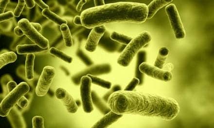
Reducing Biofilm
Biofilm is the growth of microorganisms that form on biological or artificial surfaces. Most bacteria and fungi can form biofilms that are found on the surfaces of many medical devices including urinary catheters, pacemakers, contact lenses, and ETTs.1-2 During procedures such as suctioning, bronchoscopy, or instillation of saline down the ETT, biofilm can be dislodged and travel deeper into the lungs.
With the introduction of these pathogens, there is an increase in infection and pneumonia.1 Research has found that coating the interior wall of the ETT with silver (which has a broad-spectrum antimicrobial characteristic) can reduce and delay the formation of biofilms. Removal of biofilm by scraping or shaving the interior wall of the ETT is another approach that is gaining approval. Products that accomplish this include the Ballard Liberator and Restore2 (Avanos), the CAM Rescue Cath (Omneotech), and the endOclear (Avanos).1,3-5
Biofilm formation has been found in COVID-19 patients and has been considered as one possible mechanism causing ETT occlusion. Problems with ETT occlusion associated with COVID-19 has been reported published in a case series in May 2021 where over a two month period, eleven patients experienced significant ETT blockage requiring replacement of the ETT in ten and tracheostomy in the eleventh patient.6 All of these cases had a thick, viscous material lining the inner lumen of the ETT. Based on the findings in these cases, the authors concluded that the underlying pathology was “related to an exuberant inflammatory response that occurs in patients with COVID-19 and ARDS” and not attributed to other factors such as formation of biofilm or issues with inadequate humidification.
The authors also state; “Because of the thick, highly viscous nature of secretions, we see a role for endotracheal tube cleaning devices in removing the debris…”6
Suctioning Techniques
Suctioning the airway occurs frequently in the care of intubated patients. This is accomplished by either using an open tracheal suctioning technique (OTST) or using a closed tracheal suction system (CTSS). In the OTST, the ventilator circuit is disconnected from the ETT to allow passage of a single-use disposable suction catheter. The CTSS employs a multiple-use suction catheter enclosed in a plastic sleeve that remains connected to the ETT and ventilator circuit. The CTSS allows suctioning to occur without disconnecting the ventilator circuit thus avoiding loss of PEEP, alveolar derecruitment, and hypoxemia when compared to the effects of using the OTST. The CTSS is less time-consuming and easier to use, and avoids exposing the healthcare team to airborne pathogens – which is an important consideration in the care of COVID-19 patients. In light of advantages of the CTSS, it has become a standard of care in airway management.7
Securing the ETT
Once an ETT has been placed it is important to have it secured to allow for ventilatory support, protection of the airway, enable safe suctioning, and avoid accidental extubation or migration of the ETT (from moving either too deep or too shallow in the trachea). ETTs can be secured by noncommercial approaches (cotton string, cloth tape, adhesive tape), or commercial products designed to hold the ETT and enable the tube to be repositioned from one side of the mouth to the other.8-9 There have been problems with the mechanism for securing the ETT causing decreased perfusion, pressure sores, and mucosal damage on the sides of the mouth. Periodic repositioning of the ETT has been included in airway management to avoid these issues. Studies have looked at the effectiveness of various approaches to secure the ETT including both non-commercial and commercial processes using simulated conditions (checking for pressure points, ability to withstand efforts to dislodge the ETT, etc). The results showed that no one device or approach performed well in all circumstances. However, the commercial ETT holders did allow for fairly quick and easy relocation of the tube as compared to the non-commercial approaches, which needed to be removed prior to relocation then reapplied to secure the tube.9
Intubation for COVID-19 Patients
Intubation of the trachea to place an ETT can range from fairly easy to difficult and has become much more complicated in light of COVID-19 issues. The novel virus SARS-CoV-2 is the causative agent of the disease labeled COVID-19 and is a highly contagious respiratory corona virus. Since the highest viral load of SARS-CoV-2 resides in the upper airway secretions and sputum, the intubation procedure brings greatly increased risk of exposure due to coughing, possible need for bag-mask ventilation, suctioning, and contamination of devices used to assist in intubation. Intubation is considered an aerosol-generating procedure (A-GP) and is associated with the highest risk to healthcare workers, followed by tracheostomy, non-invasive ventilation, and bag-mask ventilation.10 Other airway management activities that may be A-GP include disconnecting the ventilator circuit during use, extubation, cardiopulmonary resuscitation (prior to intubation), bronchoscopy, and suctioning using a single use disposable catheter (open tracheal suctioning technique mentioned earlier).10
Consensus guidelines from the United Kingdom, published in June 2020, provide an excellent, detailed resource regarding airway management and intubation.10 A summary of key issues covered by this document include the following:
- Consider intubation of the COVID-19 patient a high-risk procedure for those involved in the procedure and for the patient. Avoid utilizing staff who have increased risk (i.e. older age, pre-existing medical conditions, compromised immune system)
- Limit the number of members of the healthcare team – if possible have only the intubator and one assistant present with a runner outside the room
- Create COVID-19 intubation trays that can be used throughout the facility
- Wear full personal protective equipment, consider double gloving, and touch as little as possible to avoid contact with contaminated surfaces
- Use a negative pressure room with > 12 air changes/hour if possible
- Use a checklist to assist all in communication and procedure; have a printed algorithm present in the room Plan how to communicate prior to entering the room
- Prepare (as much as possible) all equipment and drugs outside the room
- Use the best skilled airway manager for the procedure to maximize first attempt success
- Pre-oxygenate with a well-fitting mask, employ a video larygoscope, bag-mask ventilate only if necessary – with two persons and use a two-handed VE-grip maximize the mask seal
- Use a supraglottic airway (i.e. laryngeal mask airway – LMA)
- Place a filtering heat-moisture exchanger (HME) at the airway connection
- Avoid aerosol-generating procedures as much as possible
- Have full monitoring in place (including continuous waveform capnography before, during, and after intubation
- Use rapid sequence induction, consider ketamine; paralyze early with rocuronium or succinylcholine and ensure full neuromuscular blockade prior to attempting intubation
- Have immediate availability of vasopressor for possible hypotension
- Intubate with a 7.0-8.0 ETT (with subglottic secretion drainage -SSD) with for females, 8.0-9.0 ETT with SSD for males
- Pass the ETT cuff 1-2 cm below the cords and inflate the cuff to seal prior to starting ventilation. Note and record the tube depth. Confirm correct placement with capnography
- Ensure all circuit connections are snug using push-twist maneuver
- Clamp ETT and pause ventilation for airway maneuvers or prior to disconnecting (bag or ventilator circuit)
- Use a standard failed intubation algorithm if difficulties arise
- Communicate clearly and calmly with repeat back, do not shout
- Insert a nasogastric tube after intubation and initiation of ventilation
- If COVID-19 status is not confirmed obtain a sputum sample for virology using a closed suction system
- Properly dispose of equipment and supplies and fully decontaminate reusable equipment following manufacturer’s instructions
- Use meticulous procedure upon leaving the room to remove PPE
- Deep clean the room 20 minutes after the procedure
- Display a record concerning ease of intubation; if it was a difficult intubation this should be communicated between shifts including a plan for how to manage possible issues (i.e. reintubation).
Conclusion
Airway management is always undergoing review and revision as new techniques, new medications, or new devices are developed to assist in this process. Novel diseases like SARS-CoV-2 may also appear that affect the procedures and precautions needed in managing the airway or dealing with other airway-related procedures. Respiratory therapists play a key role in the healthcare team and need to be up-to-date in changes or improvements related to handling this crucial aspect of care.
RT
Bill Pruitt, MBA, RRT, CPFT, AE-C, FAARC, is a writer, lecturer, and consultant and recently retired from over 20 years teaching at the University of South Alabama in Cardiorespiratory Care. He also volunteers at the Pulmonary Clinic at Victory Health Partners in Mobile, AL.
For more information, contact [email protected].
References
- Haas CF, Eakin RM, Konkle MA, Blank R. Endotracheal Tubes: Old and NewDiscussion. Respiratory care. 2014 Jun 1;59(6):933-55.
- Zhang K, Li X, Yu C, Wang Y. Promising therapeutic strategies against microbial biofilm challenges. Frontiers in Cellular and Infection Microbiology. 2020;10.
- Avanos Respiratory Health: https://avanosmedicaldevices.com/respiratory-health/ett-clearing-systems/. Accessed 8/10/2021.
- Wicker B, Salem G, Schofield L, et al. Two-year study to evaluate daily endotracheal tube cleaning protocol. SCCM Annual Meeting (San Diego, CA) February 17 19, 2019.
- Schofield L, Saurs G. The use of the endoclear catheter device to improve ventilator weaning. Chest. 2013; 144 (4_MeetingAbstracts):64A.
- Wiles S, Mireles-Cabodevila E, Neuhofs S, Mukhopadhyay S, Reynolds JP, Hatipoğlu U. Endotracheal Tube Obstruction Among Patients Mechanically Ventilated for ARDS Due to COVID-19: A Case Series. Journal of Intensive Care Medicine. 2021 May;36(5):604-11.
- Imbriaco G, Monesi A. Closed tracheal suctioning systems in the era of COVID-19: is it time to consider them as a gold standard?. Journal of Infection Prevention. 2021 Jan;22(1):44-5.
- Lovett PB, Flaxman A, Stürmann KM, Bijur P. The insecure airway: a comparison of knots and commercial devices for securing endotracheal tubes. BMC Emergency Medicine. 2006 Dec;6(1):1-7.
- Fisher DF, Chenelle CT, Marchese AD, Kratohvil JP, Kacmarek RM. “Comparison of commercial and noncommercial endotracheal tube-securing devices.” Respiratory Care Sep 2014, 59 (9) 1315-1323; DOI: 10.4187/respcare.02951
- Cook TM, El‐Boghdadly K, McGuire B, McNarry AF, Patel A, Higgs A. Consensus guidelines for managing the airway in patients with COVID‐19: Guidelines from the Difficult Airway Society, the Association of Anaesthetists the Intensive Care Society, the Faculty of Intensive Care Medicine and the Royal College of Anaesthetists. Anaesthesia. 2020 Jun;75(6):785-99.